5J76 image
Deposition Date 2016-04-06
Release Date 2016-06-22
Last Version Date 2024-11-06
Entry Detail
PDB ID:
5J76
Title:
Structure of Lectin from Colocasia esculenta(L.) Schott
Biological Source:
Source Organism:
Method Details:
Experimental Method:
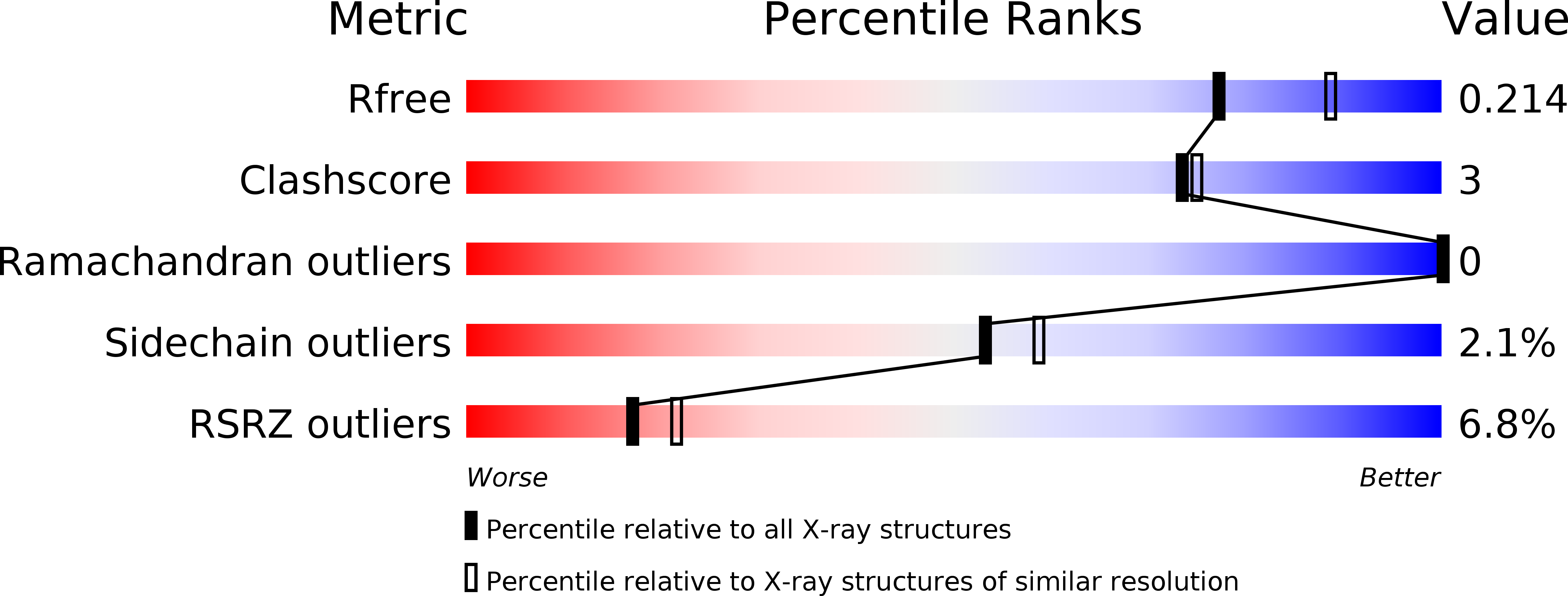
Resolution:
2.10 Å
R-Value Free:
0.20
R-Value Work:
0.16
R-Value Observed:
0.16
Space Group:
P 31 2 1
Macromolecular Entities
Polymer Type:polypeptide(L)
Molecule:12kD storage protein
Chain IDs:A
Chain Length:109
Number of Molecules:1
Biological Source:Colocasia esculenta
Polymer Type:polypeptide(L)
Molecule:12kD storage protein
Chain IDs:B
Chain Length:111
Number of Molecules:1
Biological Source:Colocasia esculenta
Ligand Molecules
Primary Citation
Structural analysis of beta-prism lectin from Colocasia esculenta (L.) S chott.
Int.J.Biol.Macromol. 91 518 523 (2016)
PMID: 27262515 DOI: 10.1016/j.ijbiomac.2016.05.048

Abstact

The Mannose-binding β-Prism Colocasia esculenta lectin (β-PCL) was purified from tubers using ion exchange chromatography. The purified β-PCL appeared as a single band of ∼12kDa on SDS-PAGE. β-PCL crystallizes in trigonal space group P3121 and diffracted to a resolution of 2.1Å. The structure was solved using Molecular replacement using Crocus vernus lectin (PDB: 3MEZ) as a model. From the final refined model to an R-factor of 16.5% and an Rfree of 20.4%, it has been observed that the biological unit consists of two β-Prism domains augmented through C-terminals swap over to form one of faces for each domain. Cα superposition of individual domains of β-PCL with individual domains of other related structures and superposition of whole protein structures were carried out. The higher RMS deviation for the superposition of whole structures suggest that β-prism domains assume different orientation in each structure.

Legend

Protein

Chemical

Disease

Primary Citation of related structures