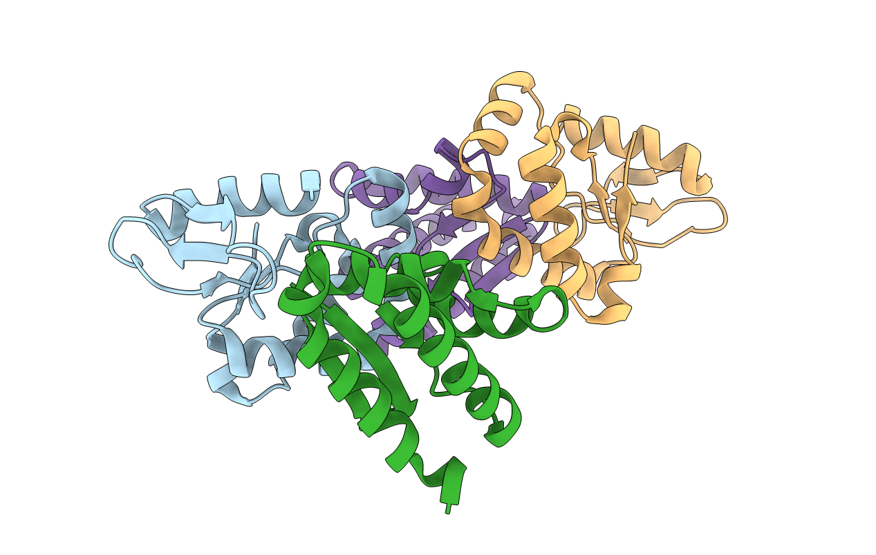
5J4A image
Entry Detail
PDB ID:
5J4A
Keywords:
Title:
CdiA-CT toxin from Burkholderia pseudomallei E479 in complex with cognate CdiI immunity protein
Biological Source:
Source Organism:
Host Organism:
PDB Version:
Deposition Date:
2016-03-31
Release Date:
2016-07-27
Method Details:
Experimental Method:
Resolution:
2.00 Å
R-Value Free:
0.23
R-Value Work:
0.19
R-Value Observed:
0.19
Space Group:
P 2 21 21
Macromolecular Entities
Polymer Type:polypeptide(L)
Description:tRNA nuclease CdiA
Mutations:D285A
Chain IDs:A, C
Chain Length:161
Number of Molecules:2
Biological Source:Burkholderia pseudomallei
Polymer Type:polypeptide(L)
Description:Immunity protein CdiI
Chain IDs:B, D
Chain Length:120
Number of Molecules:2
Biological Source:Burkholderia pseudomallei
Primary Citation
Functional Diversity of Cytotoxic tRNase/Immunity Protein Complexes from Burkholderia pseudomallei.
J.Biol.Chem. 291 19387 19400 (2016)
PMID: 27445337 DOI: 10.1074/jbc.M116.736074

Abstact

Contact-dependent growth inhibition (CDI) is a widespread mechanism of inter-bacterial competition. CDI(+) bacteria deploy large CdiA effector proteins, which carry variable C-terminal toxin domains (CdiA-CT). CDI(+) cells also produce CdiI immunity proteins that specifically neutralize cognate CdiA-CT toxins to prevent auto-inhibition. Here, we present the crystal structure of the CdiA-CT/CdiI(E479) toxin/immunity protein complex from Burkholderia pseudomallei isolate E479. The CdiA-CT(E479) tRNase domain contains a core α/β-fold that is characteristic of PD(D/E)XK superfamily nucleases. Unexpectedly, the closest structural homolog of CdiA-CT(E479) is another CDI toxin domain from B. pseudomallei 1026b. Although unrelated in sequence, the two B. pseudomallei nuclease domains share similar folds and active-site architectures. By contrast, the CdiI(E479) and CdiI(1026b) immunity proteins share no significant sequence or structural homology. CdiA-CT(E479) and CdiA-CT(1026b) are both tRNases; however, each nuclease cleaves tRNA at a distinct position. We used a molecular docking approach to model each toxin bound to tRNA substrate. The resulting models fit into electron density envelopes generated by small-angle x-ray scattering analysis of catalytically inactive toxin domains bound stably to tRNA. CdiA-CT(E479) is the third CDI toxin found to have structural homology to the PD(D/E)XK superfamily. We propose that CDI systems exploit the inherent sequence variability and active-site plasticity of PD(D/E)XK nucleases to generate toxin diversity. These findings raise the possibility that many other uncharacterized CDI toxins may belong to the PD(D/E)XK superfamily.

Legend

Protein

Chemical

Disease

Primary Citation of related structures