Deposition Date
2016-03-31
Release Date
2016-10-19
Last Version Date
2023-11-08
Entry Detail
PDB ID:
5J3R
Keywords:
Title:
Crystal structure of yeast monothiol glutaredoxin Grx6 in complex with a glutathione-coordinated [2Fe-2S] cluster
Biological Source:
Source Organism(s):
Saccharomyces cerevisiae S288c (Taxon ID: 559292)
Expression System(s):
Method Details:
Experimental Method:
Resolution:
2.46 Å
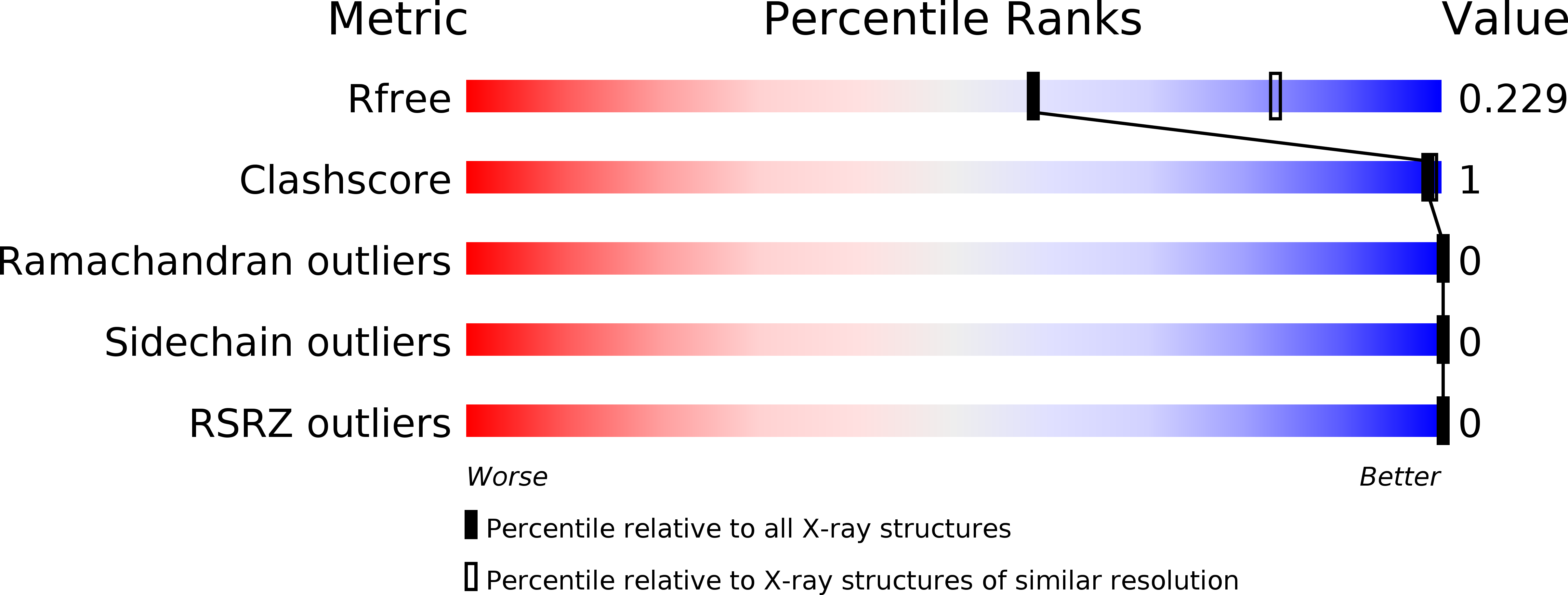
R-Value Free:
0.23
R-Value Work:
0.19
R-Value Observed:
0.19
Space Group:
P 43 21 2