5J2O image
Deposition Date 2016-03-29
Release Date 2016-04-13
Last Version Date 2024-11-13
Entry Detail
PDB ID:
5J2O
Title:
Crystal structure of the cyan fluorescence protein Cerulean S175G mutant
Biological Source:
Source Organism:
Host Organism:
Method Details:
Experimental Method:
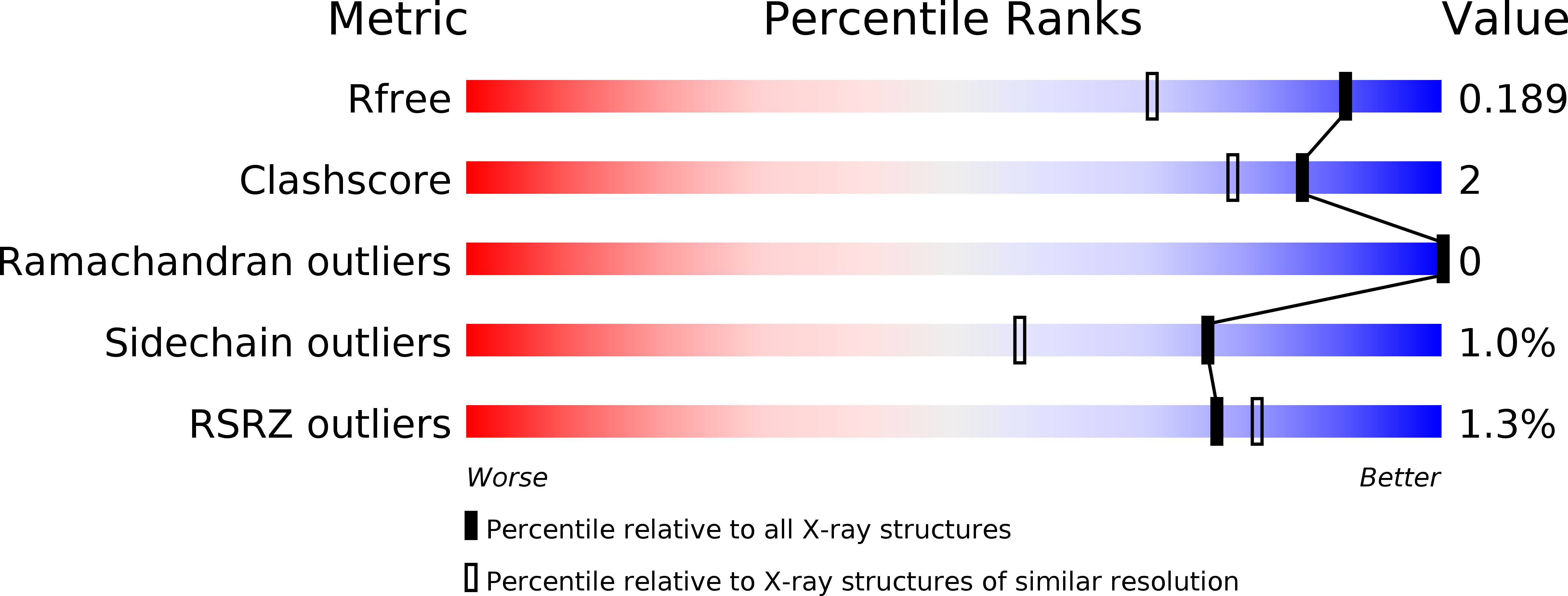
Resolution:
1.50 Å
R-Value Free:
0.18
R-Value Work:
0.16
Space Group:
P 21 21 21
Macromolecular Entities
Structures with similar UniProt ID
Protein Blast
Polymer Type:polypeptide(L)
Molecule:Green fluorescent protein
Gene (Uniprot):GFP
Mutagens:F65L/Y145A/N146I/H148D/M153T/V163A/S175G/H231L
Chain IDs:A
Chain Length:245
Number of Molecules:1
Biological Source:Aequorea victoria
Modified Residue
Compound ID Chain ID Parent Comp ID Details 2D Image
CRF A GLY chromophore
Primary Citation
Crystal structure of the cyan fluorescent protein Cerulean-S175G
Acta Crystallogr.,Sect.F 72 516 522 (2016)
PMID: 27380368 DOI: 10.1107/S2053230X16008311

Abstact

Enhanced cyan fluorescent protein (ECFP) was derived from Aequorea victoria green fluorescent protein (avGFP), notably with S65T/Y66W mutations. Its chromophore consists of a tripeptide comprised of Thr65, Trp66 and Gly67 (TWG) residues, while that of avGFP consists of a Ser65, Tyr66 and Gly67 (SYG) tripeptide. Cerulean and SCFP3A were derived from ECFP-S72A/H148D (a double mutation) with additional Y145A and S175G mutations, respectively, while Cerulean-S175G has both mutations (Y145A and S175G). The crystal structures of these ECFP variants at neutral pH were reported to adopt two distinct major conformations called ECFP and Cerulean. In this study, Cerulean-S175G was revealed to adopt only the Cerulean conformation, while Cerulean has been reported to adopt both the ECFP and the Cerulean conformations in its crystal structures. Sharing the same S175G mutation with SCFP3A, Cerulean-S175G showed a slightly increased quantum yield, like SCFP3A, but did not adopt the ECFP conformation adopted by SCFP3A. Detailed comparison of Cerulean-S175G and other ECFP variants revealed that the notable conformational changes in ECFP variants can be understood mainly in terms of the interaction between the Trp66 residue of the chromophore and residues 145-148 of β-strand 7.

Legend

Protein

Chemical

Disease

Primary Citation of related structures