Deposition Date
2016-03-28
Release Date
2017-03-15
Last Version Date
2023-09-27
Entry Detail
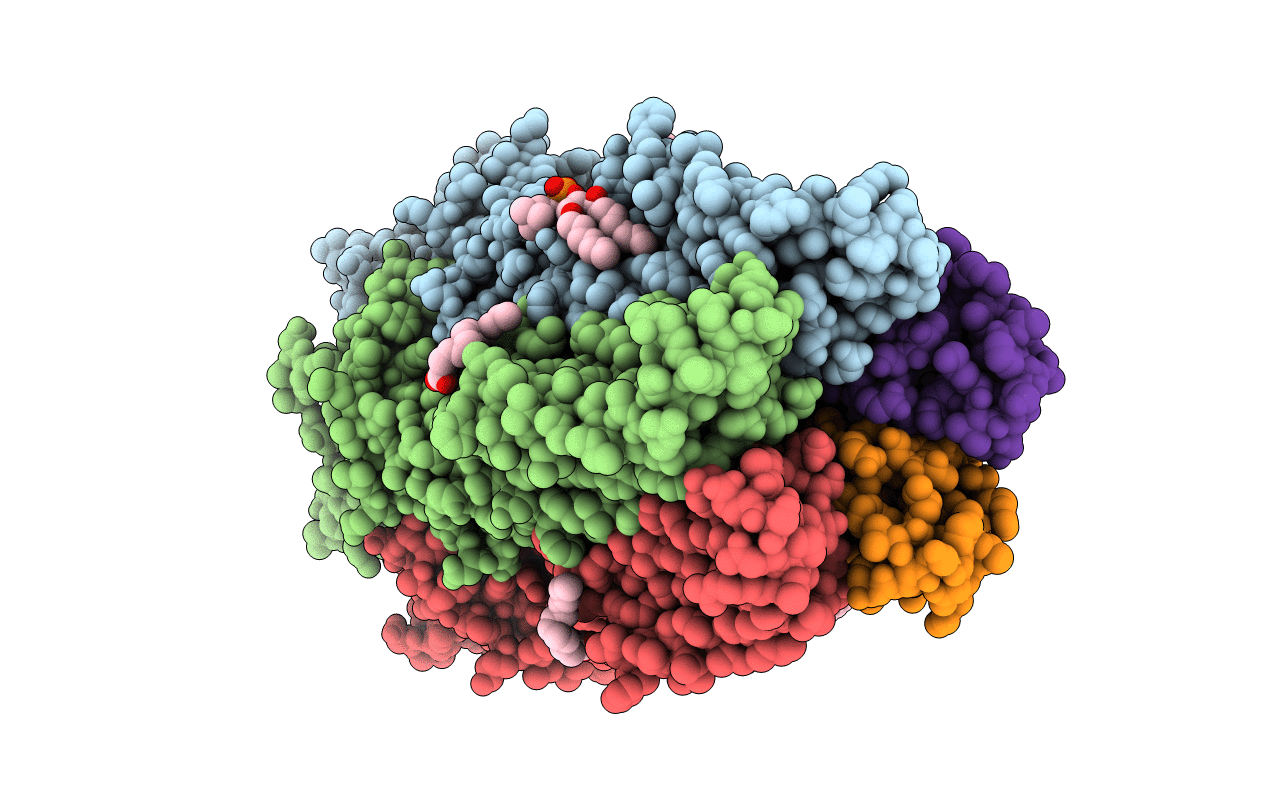
Biological Source:
Source Organism:
Gloeobacter violaceus (strain PCC 7421) (Taxon ID: 251221)
Host Organism:
Method Details:
Experimental Method:
Resolution:
3.25 Å
R-Value Free:
0.26
R-Value Work:
0.23
R-Value Observed:
0.23
Space Group:
C 1 2 1