
Deposition Date
2016-03-17
Release Date
2017-04-05
Last Version Date
2024-05-08
Entry Detail
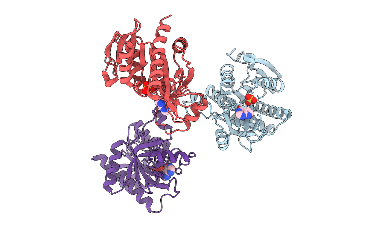
PDB ID:
5IU6
Keywords:
Title:
Crystal structure of E.coli purine nucleoside phosphorylase with 7-deazahypoxanthine
Biological Source:
Source Organism(s):
Escherichia coli (Taxon ID: 562)
Expression System(s):
Method Details:
Experimental Method:
Resolution:
2.51 Å
R-Value Free:
0.20
R-Value Work:
0.15
R-Value Observed:
0.15
Space Group:
P 61 2 2
