5IRU image
Deposition Date 2016-03-14
Release Date 2016-10-12
Last Version Date 2024-10-16
Entry Detail
PDB ID:
5IRU
Title:
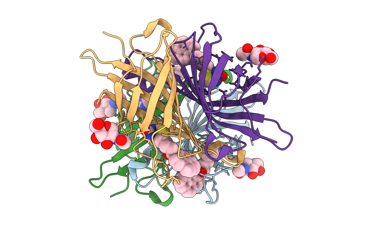
Crystal structure of avidin in complex with 1-biotinylpyrene
Biological Source:
Source Organism:
Gallus gallus (Taxon ID: 9031)
Method Details:
Experimental Method:
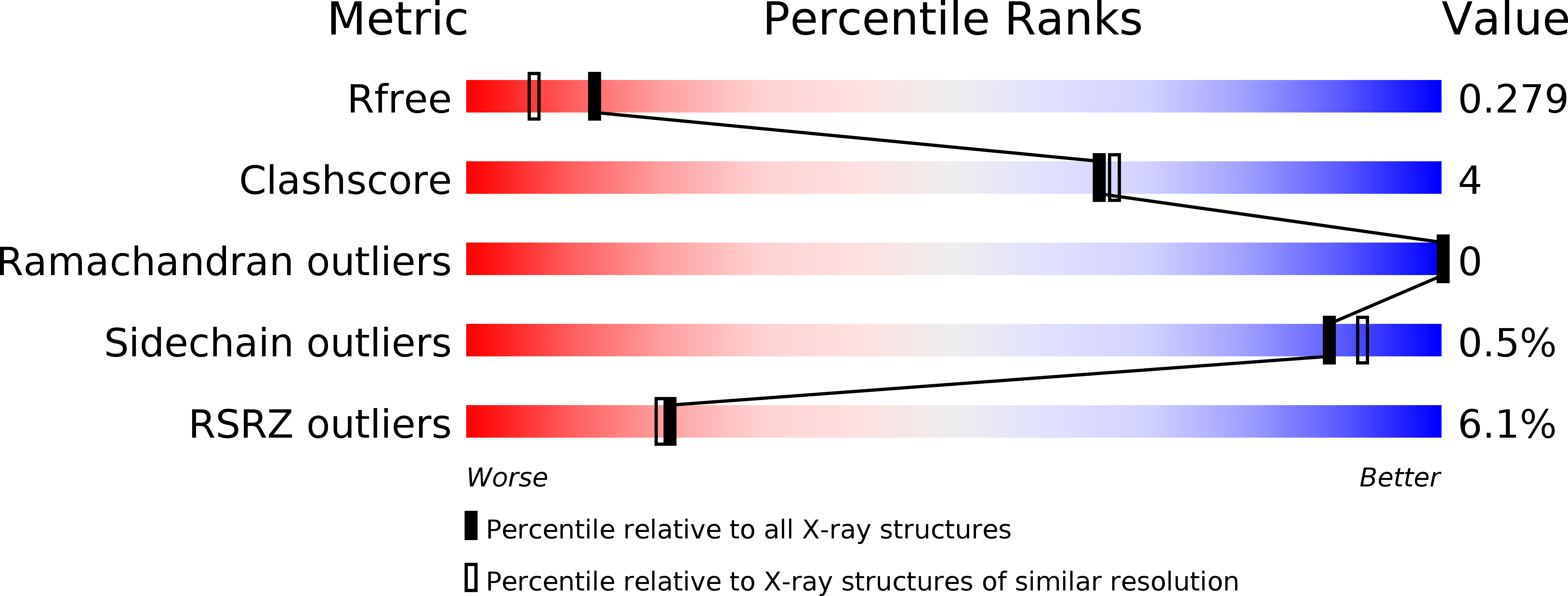
Resolution:
2.00 Å
R-Value Free:
0.26
R-Value Work:
0.22
R-Value Observed:
0.22
Space Group:
P 21 21 21
Macromolecular Entities
Polymer Type:polypeptide(L)
Molecule:Avidin
Gene (Uniprot):AVD
Chain IDs:A, B, C, D
Chain Length:128
Number of Molecules:4
Biological Source:Gallus gallus
Primary Citation
Structural Characterization of the Avidin Interactions with Fluorescent Pyrene-Conjugates: 1-Biotinylpyrene and 1-Desthiobiotinylpyrene.
Molecules 21 ? ? (2016)
PMID: 27689976 DOI: 10.3390/molecules21101270

Abstact

Avidin is a tetrameric protein that belongs to the calycin superfamily. It has been studied mainly because of its extraordinary affinity to biotin, which led to a wide range of applications based on the avidin-biotin system. In the present study, we report the first crystal structures of avidin in a complex with two novel fluorescent pyrene derivatives: 1-biotinylpyrene (B9P) and 1-desthiobiotinylpyrene (D9P). The crystal structures were solved by molecular replacement using the coordinates of avidin molecule as a starting model and the final models of avidin/B9P and avidin/D9P were refined to resolutions of 2.0 Å and 2.1 Å, respectively. Our data reveal changes in loop conformation as well as in overall fold and quaternary arrangement of the avidin upon the binding of these fluorescent probes. Moreover, the crystal structures allowed analysis of the details of the interactions between the protein and the pyrene derivatives. Structural description of the complexes will contribute to the design of conjugates for expanding the capabilities of avidin-biotin technology.

Legend

Protein

Chemical

Disease

Primary Citation of related structures