5IR6 image
Entry Detail
PDB ID:
5IR6
Keywords:
Title:
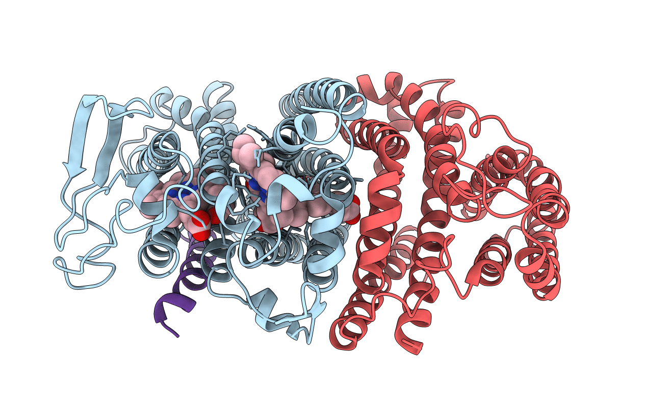
The structure of bd oxidase from Geobacillus thermodenitrificans
Biological Source:
PDB Version:
Deposition Date:
2016-03-12
Release Date:
2016-05-04
Method Details:
Experimental Method:
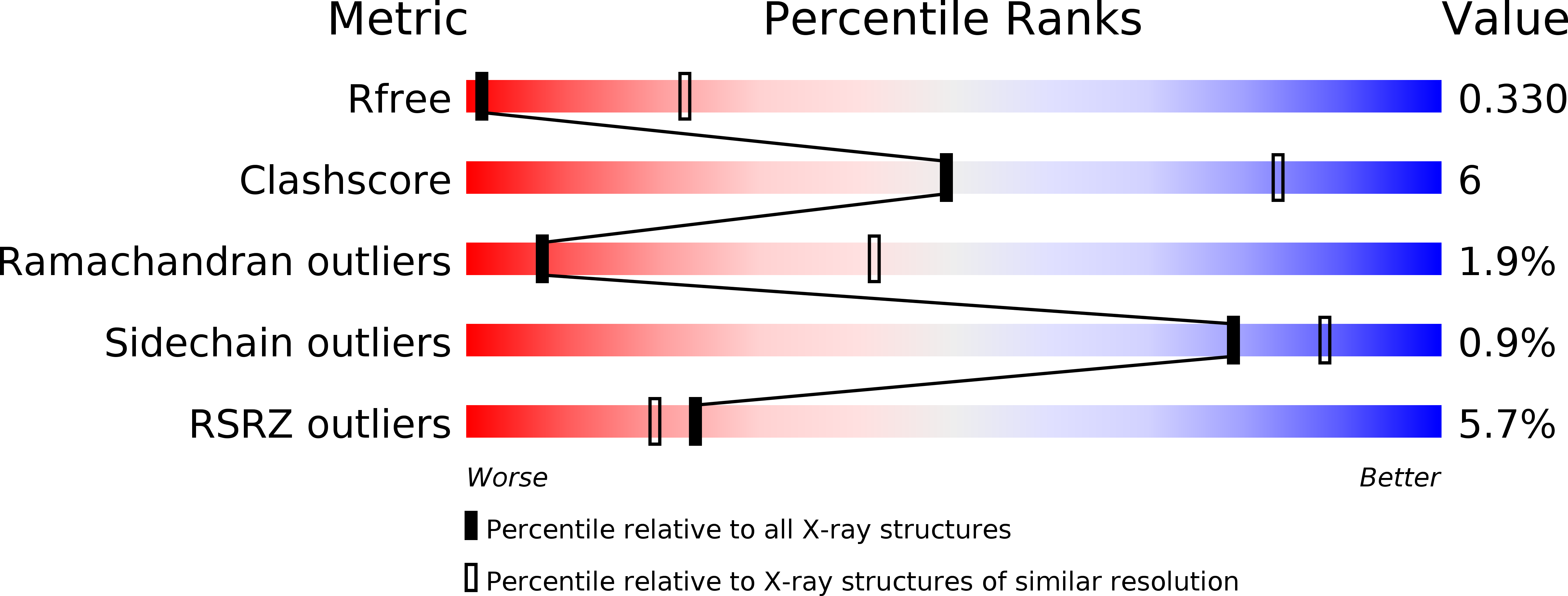
Resolution:
3.80 Å
R-Value Free:
0.32
R-Value Work:
0.30
R-Value Observed:
0.30
Space Group:
P 21 21 21
Macromolecular Entities
Polymer Type:polypeptide(L)
Description:Bd-type quinol oxidase subunit I
Chain IDs:A
Chain Length:448
Number of Molecules:1
Biological Source:Geobacillus stearothermophilus K1041
Polymer Type:polypeptide(L)
Description:Bd-type quinol oxidase subunit II
Chain IDs:B
Chain Length:342
Number of Molecules:1
Biological Source:Geobacillus stearothermophilus K1041
Polymer Type:polypeptide(L)
Description:Putative membrane protein
Chain IDs:C
Chain Length:33
Number of Molecules:1
Biological Source:Geobacillus sp. PA-3
Primary Citation
Structure of a bd oxidase indicates similar mechanisms for membrane-integrated oxygen reductases.
Science 352 583 586 (2016)
PMID: 27126043 DOI: 10.1126/science.aaf2477

Abstact

The cytochrome bd oxidases are terminal oxidases that are present in bacteria and archaea. They reduce molecular oxygen (dioxygen) to water, avoiding the production of reactive oxygen species. In addition to their contribution to the proton motive force, they mediate viability under oxygen-related stress conditions and confer tolerance to nitric oxide, thus contributing to the virulence of pathogenic bacteria. Here we present the atomic structure of the bd oxidase from Geobacillus thermodenitrificans, revealing a pseudosymmetrical subunit fold. The arrangement and order of the heme cofactors support the conclusions from spectroscopic measurements that the cleavage of the dioxygen bond may be mechanistically similar to that in the heme-copper-containing oxidases, even though the structures are completely different.

Legend

Protein

Chemical

Disease

Primary Citation of related structures