Deposition Date
2016-03-03
Release Date
2016-08-24
Last Version Date
2024-01-10
Entry Detail
PDB ID:
5IKB
Keywords:
Title:
Crystal structure of the kainate receptor GluK4 ligand binding domain in complex with kainate
Biological Source:
Source Organism(s):
Rattus norvegicus (Taxon ID: 10116)
Expression System(s):
Method Details:
Experimental Method:
Resolution:
2.05 Å
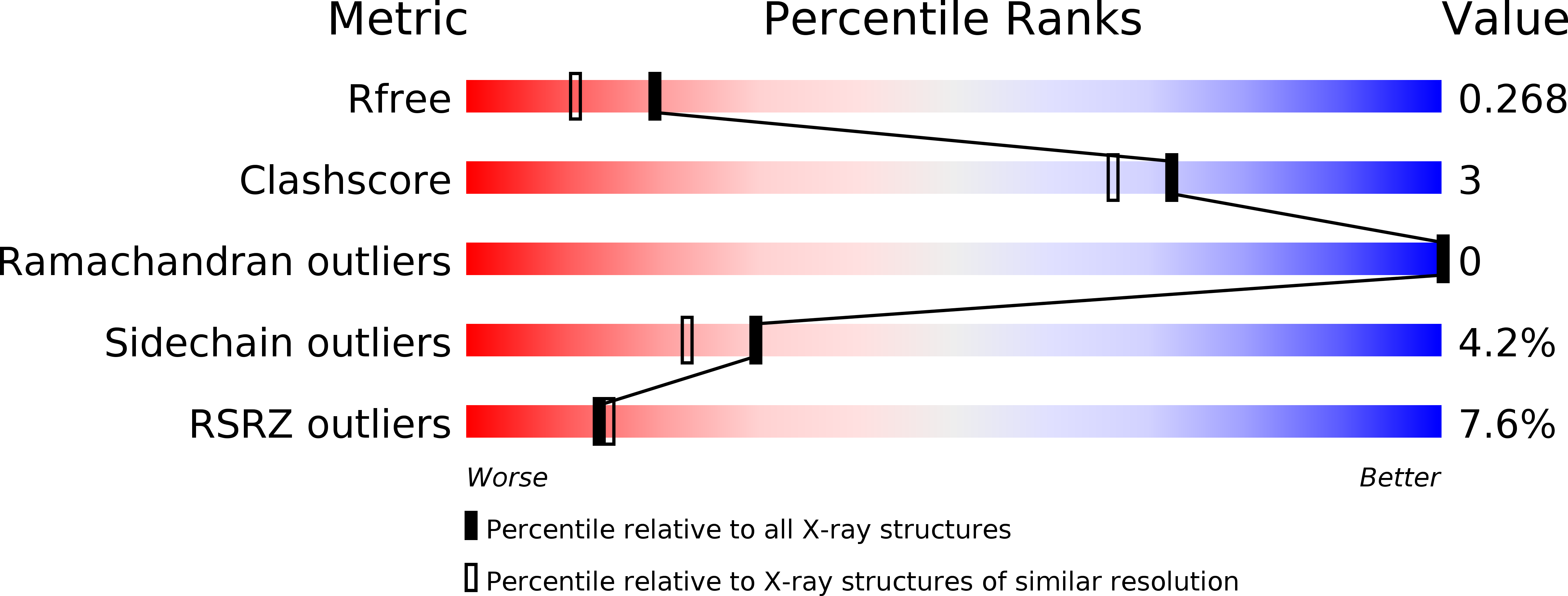
R-Value Free:
0.26
R-Value Work:
0.19
R-Value Observed:
0.19
Space Group:
C 2 2 21