5IK7 image
Entry Detail
PDB ID:
5IK7
Title:
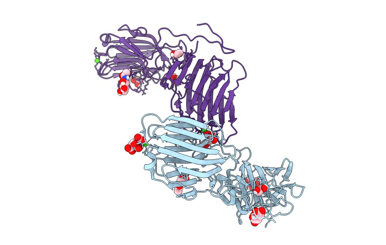
Laminin A2LG45 I-form, Apo.
Biological Source:
Source Organism:
Host Organism:
PDB Version:
Deposition Date:
2016-03-03
Release Date:
2016-08-10
Method Details:
Experimental Method:
Resolution:
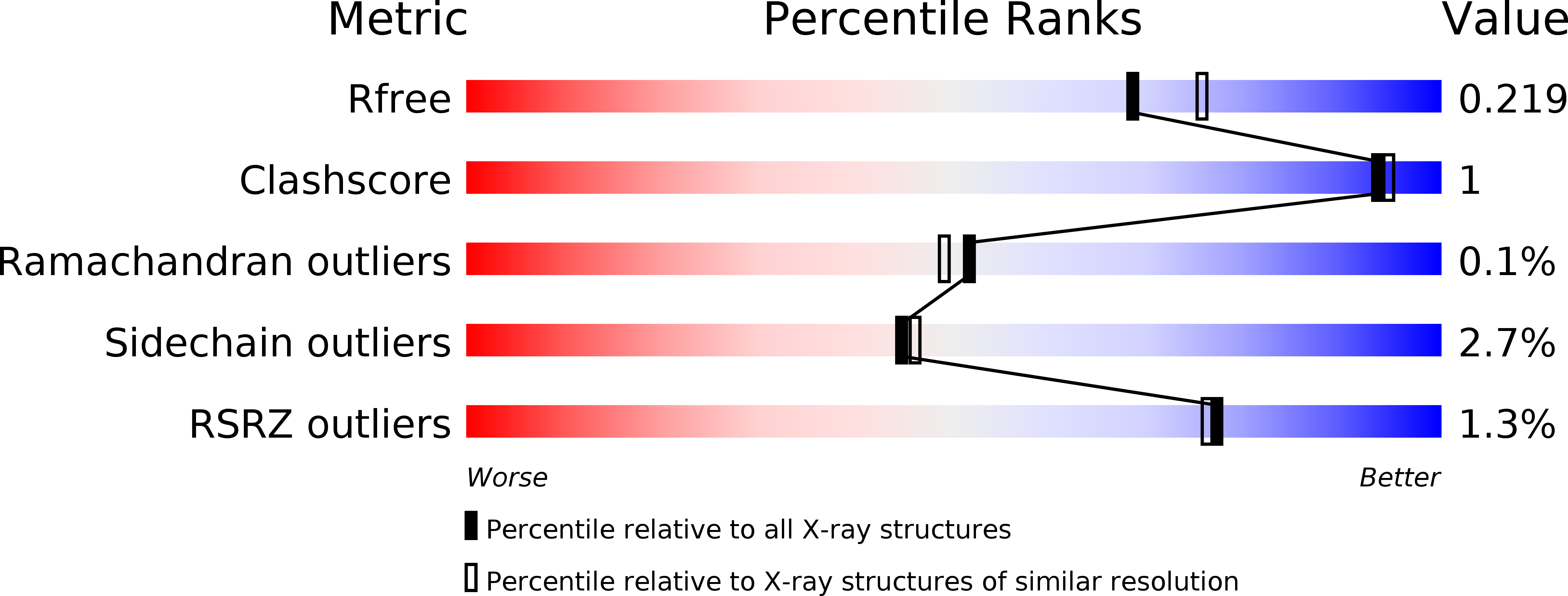
2.00 Å
R-Value Free:
0.21
R-Value Work:
0.18
R-Value Observed:
0.18
Space Group:
I 21 21 21
Macromolecular Entities
Polymer Type:polypeptide(L)
Description:Laminin subunit alpha-2
Chain IDs:A, B
Chain Length:381
Number of Molecules:2
Biological Source:Mus musculus
Primary Citation
Structural basis of laminin binding to the LARGE glycans on dystroglycan.
Nat.Chem.Biol. 12 810 814 (2016)
PMID: 27526028 DOI: 10.1038/nchembio.2146

Abstact

Dystroglycan is a highly glycosylated extracellular matrix receptor with essential functions in skeletal muscle and the nervous system. Reduced matrix binding by α-dystroglycan (α-DG) due to perturbed glycosylation is a pathological feature of several forms of muscular dystrophy. Like-acetylglucosaminyltransferase (LARGE) synthesizes the matrix-binding heteropolysaccharide [-glucuronic acid-β1,3-xylose-α1,3-]n. Using a dual exoglycosidase digestion, we confirm that this polysaccharide is present on native α-DG from skeletal muscle. The atomic details of matrix binding were revealed by a high-resolution crystal structure of laminin-G-like (LG) domains 4 and 5 (LG4 and LG5) of laminin-α2 bound to a LARGE-synthesized oligosaccharide. A single glucuronic acid-β1,3-xylose disaccharide repeat straddles a Ca(2+) ion in the LG4 domain, with oxygen atoms from both sugars replacing Ca(2+)-bound water molecules. The chelating binding mode accounts for the high affinity of this protein-carbohydrate interaction. These results reveal a previously uncharacterized mechanism of carbohydrate recognition and provide a structural framework for elucidating the mechanisms underlying muscular dystrophy.

Legend

Protein

Chemical

Disease

Primary Citation of related structures