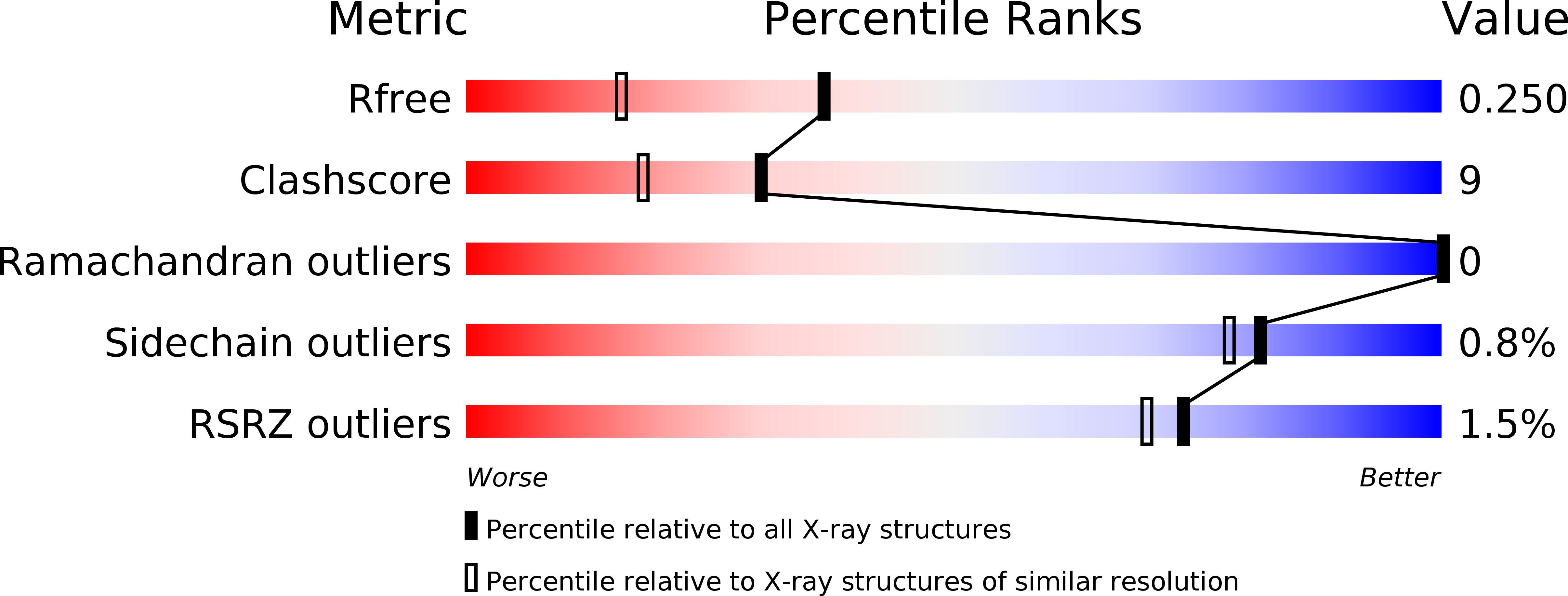
5IH2 image
Deposition Date 2016-02-28
Release Date 2016-06-29
Last Version Date 2024-10-30
Entry Detail
PDB ID:
5IH2
Title:
Structure, thermodynamics, and the role of conformational dynamics in the interactions between the N-terminal SH3 domain of CrkII and proline-rich motifs in cAbl
Biological Source:
Source Organism:
Mus musculus (Taxon ID: 10090)
Endothia gyrosa (Taxon ID: 40263)
Host Organism:
Method Details:
Experimental Method:
Resolution:
1.80 Å
R-Value Free:
0.24
R-Value Work:
0.16
R-Value Observed:
0.17
Space Group:
P 1 21 1
Macromolecular Entities
Polymer Type:polypeptide(L)
Molecule:Adapter molecule crk
Gene (Uniprot):Crk
Chain IDs:A, B
Chain Length:58
Number of Molecules:2
Biological Source:Mus musculus
Polymer Type:polypeptide(L)
Molecule:Proline rich Peptide
Chain IDs:C (auth: M), D (auth: N)
Chain Length:12
Number of Molecules:2
Biological Source:Endothia gyrosa
Primary Citation
Binding Mechanism of the N-Terminal SH3 Domain of CrkII and Proline-Rich Motifs in cAbl.
Biophys.J. 110 2630 2641 (2016)
PMID: 27332121 DOI: 10.1016/j.bpj.2016.05.008

Abstact

The N-terminal Src homology 3 (nSH3) domain of a signaling adaptor protein, CT-10 regulator of kinase II (CrkII), recognizes proline-rich motifs (PRMs) of binding partners, such as cAbl kinase. The interaction between CrkII and cAbl kinase is involved in the regulation of cell spreading, microbial pathogenesis, and cancer metastasis. Here, we report the detailed biophysical characterizations of the interactions between the nSH3 domain of CrkII and PRMs in cAbl. We identified that the nSH3 domain of CrkII binds to three PRMs in cAbl with virtually identical affinities. Structural studies, by using x-ray crystallography and NMR spectroscopy, revealed that the binding modes of all three nSH3:PRM complexes are highly similar to each other. Van 't Hoff analysis revealed that nSH3:PRM interaction is associated with favorable enthalpy and unfavorable entropy change. The combination of experimentally determined thermodynamic parameters, structure-based calculations, and (15)N NMR relaxation analysis highlights the energetic contribution of conformational entropy change upon the complex formation, and water molecules structured in the binding interface of the nSH3:PRM complex. Understanding the molecular basis of nSH3:PRM interaction will provide, to our knowledge, new insights for the rational design of small molecules targeting the interaction between CrkII and cAbl.

Legend

Protein

Chemical

Disease

Primary Citation of related structures