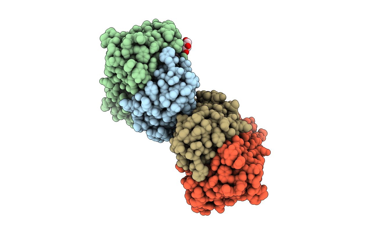
5ID0 image
Entry Detail
PDB ID:
5ID0
Keywords:
Title:
Cetuximab Fab in complex with aminoheptanoic acid-linked meditope
Biological Source:
PDB Version:
Deposition Date:
2016-02-23
Release Date:
2016-06-15
Method Details:
Experimental Method:
Resolution:
2.48 Å
R-Value Free:
0.20
R-Value Work:
0.16
R-Value Observed:
0.16
Space Group:
P 21 21 21
Macromolecular Entities
Polymer Type:polypeptide(L)
Description:Cetuximab Fab light chain
Chain IDs:A, C
Chain Length:213
Number of Molecules:2
Biological Source:MUS MUSCULUS, HOMO SAPIENS
Polymer Type:polypeptide(L)
Description:Cetuximab Fab heavy chain
Chain IDs:B, D
Chain Length:221
Number of Molecules:2
Biological Source:MUS MUSCULUS, HOMO SAPIENS
Polymer Type:polypeptide(L)
Description:Cyclic meditope
Chain IDs:E, F
Chain Length:11
Number of Molecules:2
Biological Source:synthetic construct
Modified Residue
Compound ID Chain ID Parent Comp ID Details 2D Image
ASN B ASN GLYCOSYLATION SITE
PCA B GLN PYROGLUTAMIC ACID
Primary Citation
Cyclization strategies of meditopes: affinity and diffraction studies of meditope-Fab complexes.
Acta Crystallogr F Struct Biol Commun 72 434 442 (2016)
PMID: 27303895 DOI: 10.1107/S2053230X16007202

Abstact

Recently, a unique binding site for a cyclic 12-residue peptide was discovered within a cavity formed by the light and heavy chains of the cetuximab Fab domain. In order to better understand the interactions that drive this unique complex, a number of variants including the residues within the meditope peptide and the antibody, as well as the cyclization region of the meditope peptide, were created. Here, multiple crystal structures of meditope peptides incorporating different cyclization strategies bound to the central cavity of the cetuximab Fab domain are presented. The affinity of each cyclic derivative for the Fab was determined by surface plasmon resonance and correlated to structural differences. Overall, it was observed that the disulfide bond used to cyclize the peptide favorably packs against a hydrophobic `pocket' and that amidation and acetylation of the original disulfide meditope increased the overall affinity ∼2.3-fold. Conversely, replacing the terminal cysteines with serines and thus creating a linear peptide reduced the affinity over 50-fold, with much of this difference being reflected in a decrease in the on-rate. Other cyclization methods, including the formation of a lactam, reduced the affinity but not to the extent of the linear peptide. Collectively, the structural and kinetic data presented here indicate that small perturbations introduced by different cyclization strategies can significantly affect the affinity of the meditope-Fab complex.

Legend

Protein

Chemical

Disease

Primary Citation of related structures