5I9E image
Deposition Date 2016-02-20
Release Date 2016-07-27
Last Version Date 2023-11-08
Entry Detail
PDB ID:
5I9E
Keywords:
Title:
Crystal structure of a nuclear actin ternary complex
Biological Source:
Method Details:
Experimental Method:
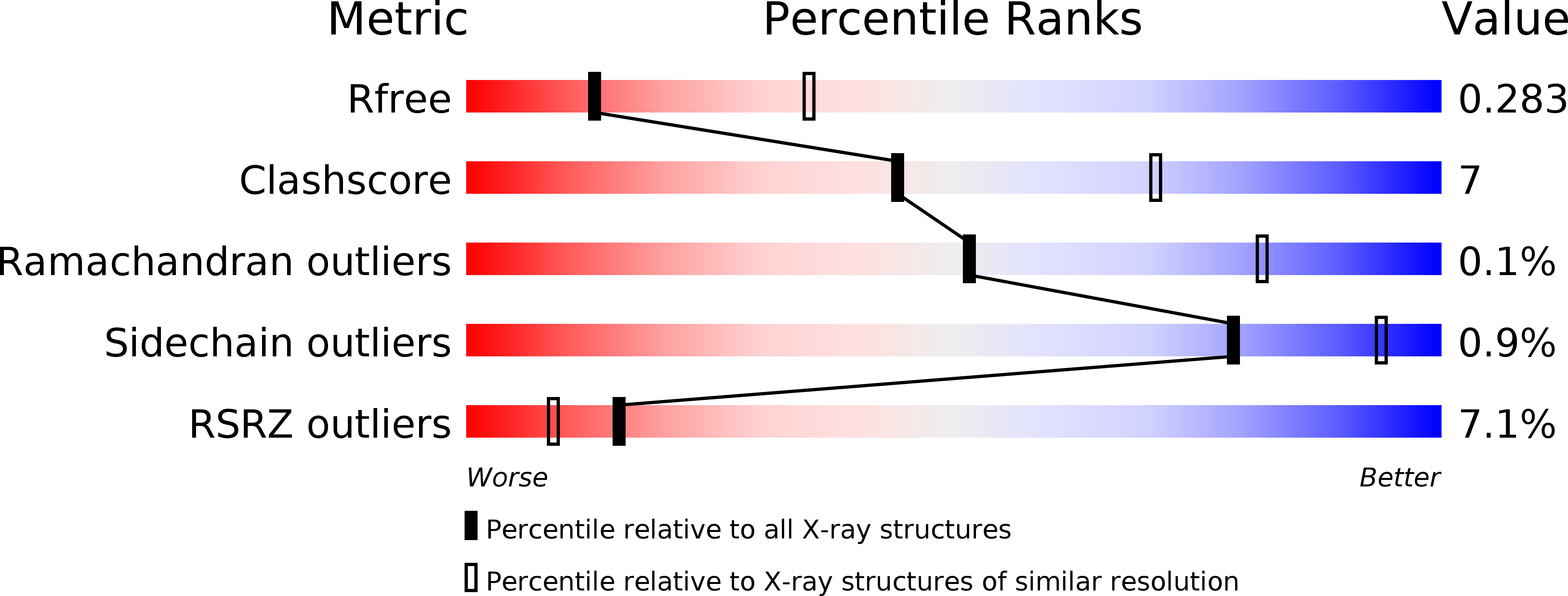
Resolution:
2.80 Å
R-Value Free:
0.28
R-Value Work:
0.23
R-Value Observed:
0.23
Space Group:
P 21 21 2
Macromolecular Entities
Polymer Type:polypeptide(L)
Molecule:Actin-related protein 4
Gene (Uniprot):ARP4
Chain IDs:A, C
Chain Length:490
Number of Molecules:2
Biological Source:Saccharomyces cerevisiae (strain ATCC 204508 / S288c)
Polymer Type:polypeptide(L)
Molecule:Actin
Gene (Uniprot):ACT1
Chain IDs:B, D
Chain Length:375
Number of Molecules:2
Biological Source:Saccharomyces bayanus
Polymer Type:polypeptide(L)
Molecule:Helicase SWR1
Gene (Uniprot):SWR1
Chain IDs:E (auth: H), F (auth: E)
Chain Length:77
Number of Molecules:2
Biological Source:Saccharomyces cerevisiae (strain ATCC 204508 / S288c)
Primary Citation
Crystal structure of a nuclear actin ternary complex.
Proc.Natl.Acad.Sci.USA 113 8985 8990 (2016)
PMID: 27457955 DOI: 10.1073/pnas.1602818113

Abstact

Actin polymerizes and forms filamentous structures (F-actin) in the cytoplasm of eukaryotic cells. It also exists in the nucleus and regulates various nucleic acid transactions, particularly through its incorporation into multiple chromatin-remodeling complexes. However, the specific structure of actin and the mechanisms that regulate its polymeric nature inside the nucleus remain unknown. Here, we report the crystal structure of nuclear actin (N-actin) complexed with actin-related protein 4 (Arp4) and the helicase-SANT-associated (HSA) domain of the chromatin remodeler Swr1. The inner face and barbed end of N-actin are sequestered by interactions with Arp4 and the HSA domain, respectively, which prevents N-actin from polymerization and binding to many actin regulators. The two major domains of N-actin are more twisted than those of globular actin (G-actin), and its nucleotide-binding pocket is occluded, freeing N-actin from binding to and regulation by ATP. These findings revealed the salient structural features of N-actin that distinguish it from its cytoplasmic counterpart and provide a rational basis for its functions and regulation inside the nucleus.

Legend

Protein

Chemical

Disease

Primary Citation of related structures