5I77 image
Deposition Date 2016-02-17
Release Date 2016-12-21
Last Version Date 2024-11-13
Entry Detail
PDB ID:
5I77
Keywords:
Title:
Crystal structure of a beta-1,4-endoglucanase from Aspergillus niger
Biological Source:
Source Organism:
Host Organism:
Method Details:
Experimental Method:
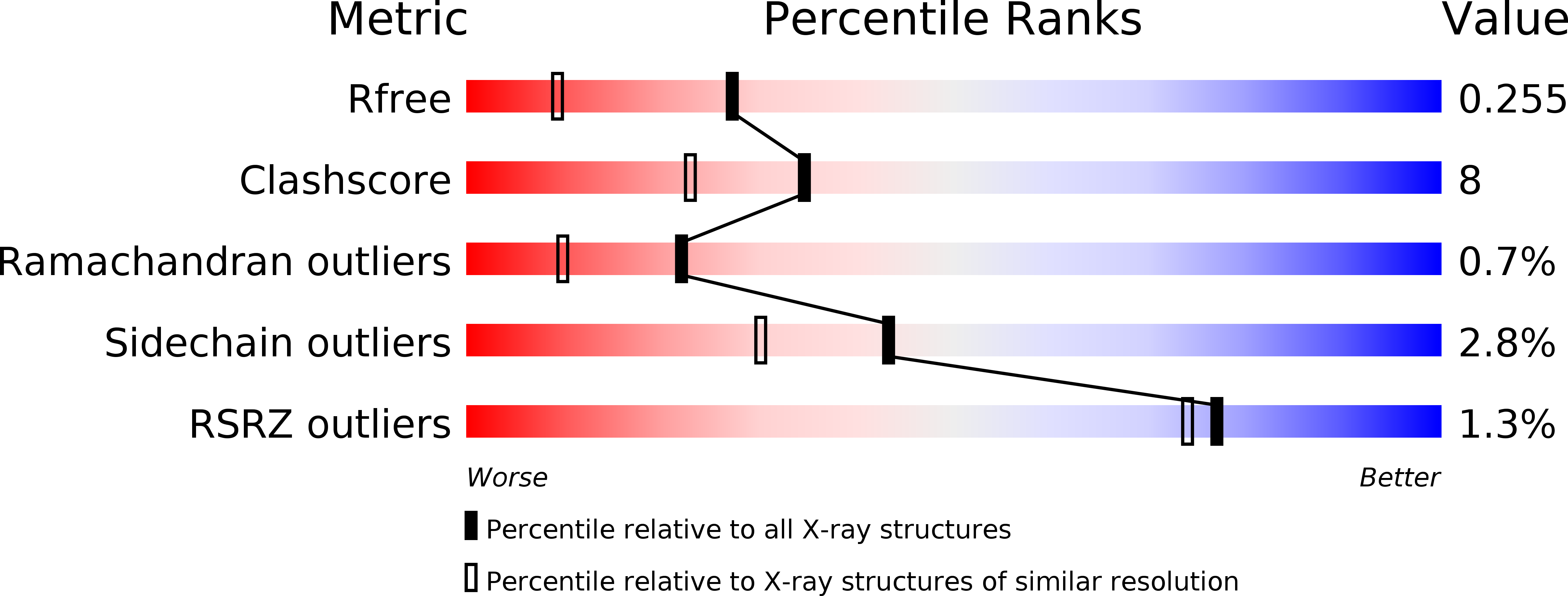
Resolution:
1.80 Å
R-Value Free:
0.24
R-Value Work:
0.18
R-Value Observed:
0.18
Space Group:
P 41 21 2
Macromolecular Entities
Structures with similar UniProt ID
Protein Blast
Polymer Type:polypeptide(L)
Molecule:Endo-beta-1, 4-glucanase
Chain IDs:A
Chain Length:302
Number of Molecules:1
Biological Source:Aspergillus niger
Primary Citation
Functional and structural analysis of Pichia pastoris-expressed Aspergillus niger 1,4-beta-endoglucanase
Biochem. Biophys. Res. Commun. 475 8 12 (2016)
PMID: 27154222 DOI: 10.1016/j.bbrc.2016.05.012

Abstact

Eukaryotic 1,4-β-endoglucanases (EC 3.2.1.4) have shown great potentials in many commercial applications because they effectively catalyze hydrolysis of cellulose, the main component of the plant cell wall. Here we expressed a glycoside hydrolase family (GH) 5 1,4-β-endoglucanase from Aspergillus niger (AnCel5A) in Pichia pastoris, which exhibits outstanding pH and heat stability. In order to further investigate the molecular mechanism of AnCel5A, apo-form and cellotetraose (CTT) complex enzyme crystal structures were solved to high resolution. AnCel5A folds into a typical (β/α)8-TIM barrel architecture, resembling other GH5 members. In the substrate binding cavity, CTT is found to bind to -4 - -1 subsites, and several polyethylene glycol molecules are found in positive subsites. In addition, several unique N-glycosylation motifs that may contribute to protein higher stability were observed from crystal structures. These results are of great importance for understanding the molecular mechanism of AnCel5A, and also provide guidance for further applications of the enzyme.

Legend

Protein

Chemical

Disease

Primary Citation of related structures