5I42 image
Deposition Date 2016-02-11
Release Date 2016-06-01
Last Version Date 2023-09-27
Entry Detail
PDB ID:
5I42
Keywords:
Title:
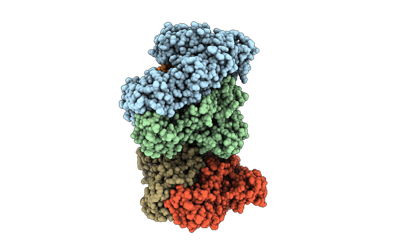
Structure of HIV-1 Reverse Transcriptase in complex with a DNA aptamer, AZTTP, and CA(2+) ion
Biological Source:
Method Details:
Experimental Method:
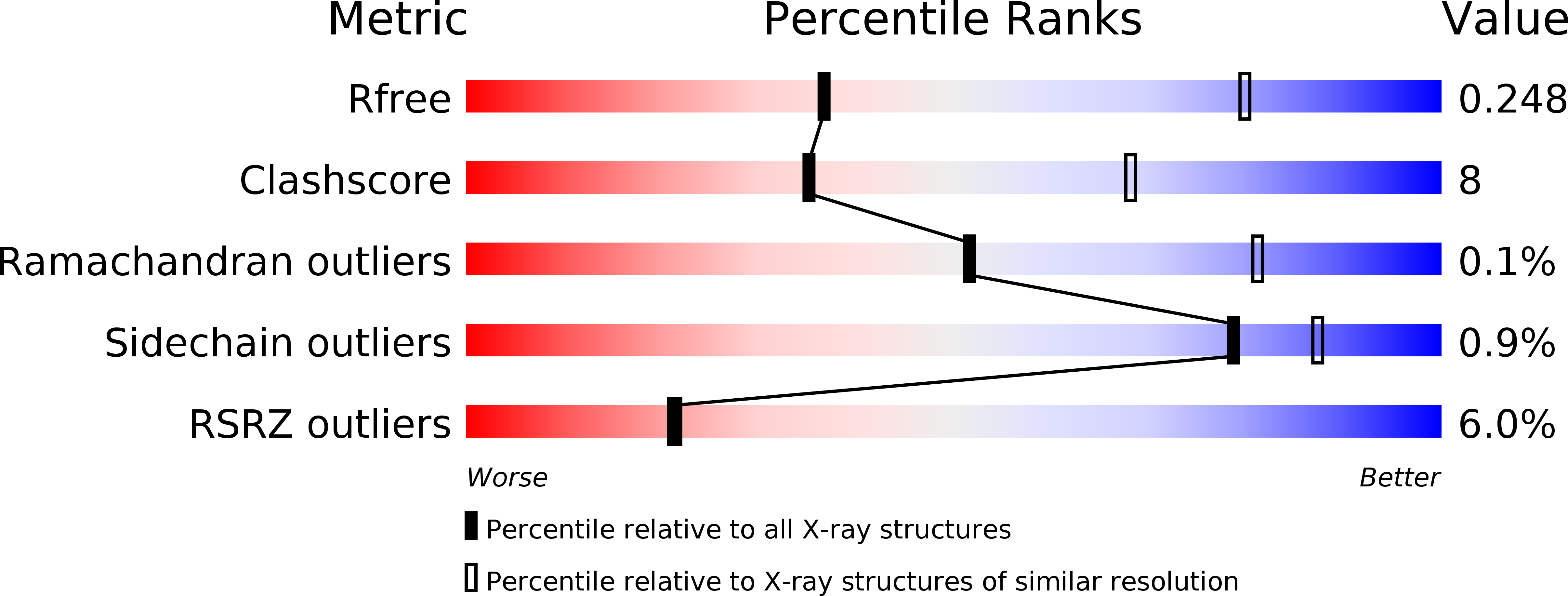
Resolution:
3.30 Å
R-Value Free:
0.24
R-Value Work:
0.22
R-Value Observed:
0.22
Space Group:
P 1 21 1
Macromolecular Entities
Polymer Type:polypeptide(L)
Molecule:HIV-1 REVERSE TRANSCRIPTASE P66 SUBUNIT
Gene (Uniprot):gag-pol
Chain IDs:A, C
Chain Length:555
Number of Molecules:2
Biological Source:Human immunodeficiency virus type 1 group M subtype B (isolate BH10)
Polymer Type:polypeptide(L)
Molecule:HIV-1 REVERSE TRANSCRIPTASE P51 SUBUNIT
Gene (Uniprot):gag-pol
Chain IDs:B, D
Chain Length:444
Number of Molecules:2
Biological Source:Human immunodeficiency virus type 1 group M subtype B (isolate BH10)
Polymer Type:polydeoxyribonucleotide
Molecule:DNA (38-MER)
Chain IDs:E, F
Chain Length:38
Number of Molecules:2
Biological Source:synthetic construct
Primary Citation
Conformational States of HIV-1 Reverse Transcriptase for Nucleotide Incorporation vs Pyrophosphorolysis-Binding of Foscarnet.
Acs Chem.Biol. 11 2158 2164 (2016)
PMID: 27192549 DOI: 10.1021/acschembio.6b00187

Abstact

HIV-1 reverse transcriptase (RT) catalytically incorporates individual nucleotides into a viral DNA strand complementing an RNA or DNA template strand; the polymerase active site of RT adopts multiple conformational and structural states while performing this task. The states associated are dNTP binding at the N site, catalytic incorporation of a nucleotide, release of a pyrophosphate, and translocation of the primer 3'-end to the P site. Structural characterization of each of these states may help in understanding the molecular mechanisms of drug activity and resistance and in developing new RT inhibitors. Using a 38-mer DNA template-primer aptamer as the substrate mimic, we crystallized an RT/dsDNA complex that is catalytically active, yet translocation-incompetent in crystals. The ability of RT to perform dNTP binding and incorporation in crystals permitted obtaining a series of structures: (I) RT/DNA (P-site), (II) RT/DNA/AZTTP ternary, (III) RT/AZT-terminated DNA (N-site), and (IV) RT/AZT-terminated DNA (N-site)/foscarnet complexes. The stable N-site complex permitted the binding of foscarnet as a pyrophosphate mimic. The Mg(2+) ions dissociated after catalytic addition of AZTMP in the pretranslocated structure III, whereas ions A and B had re-entered the active site to bind foscarnet in structure IV. The binding of foscarnet involves chelation with the Mg(2+) (B) ion and interactions with K65 and R72. The analysis of interactions of foscarnet and the recently discovered nucleotide-competing RT inhibitor (NcRTI) α-T-CNP in two different conformational states of the enzyme provides insights for developing new classes of polymerase active site RT inhibitors.

Legend

Protein

Chemical

Disease

Primary Citation of related structures