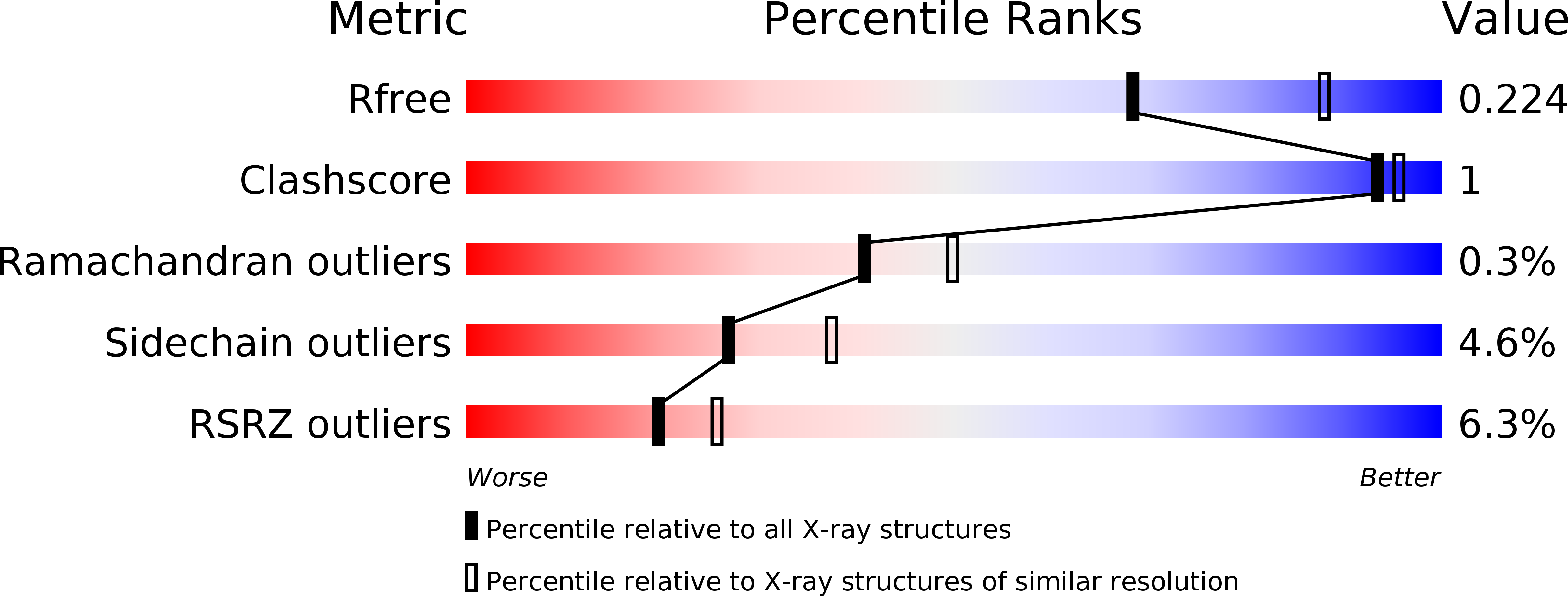
5I3C image
Deposition Date 2016-02-10
Release Date 2017-02-22
Last Version Date 2024-01-10
Entry Detail
PDB ID:
5I3C
Keywords:
Title:
Crystal structure of E.coli purine nucleoside phosphorylase with acycloguanosine
Biological Source:
Host Organism:
Method Details:
Experimental Method:
Resolution:
2.32 Å
R-Value Free:
0.21
R-Value Work:
0.17
R-Value Observed:
0.17
Space Group:
P 61 2 2
Macromolecular Entities
Polymer Type:polypeptide(L)
Molecule:Purine nucleoside phosphorylase DeoD-type
Gene (Uniprot):deoD
Chain IDs:A, B, C
Chain Length:237
Number of Molecules:3
Biological Source:Escherichia coli O139:H28 (strain E24377A / ETEC)
Primary Citation
Crystal structure of Escherichia coli purine nucleoside phosphorylase complexed with acyclovir.
Acta Crystallogr F Struct Biol Commun 74 402 409 (2018)
PMID: 29969103 DOI: 10.1107/S2053230X18008087

Abstact

Escherichia coli purine nucleoside phosphorylase (PNP), which catalyzes the reversible phosphorolysis of purine ribonucleosides, belongs to the family I hexameric PNPs. Owing to their key role in the purine salvage pathway, PNPs are attractive targets for drug design against some pathogens. Acyclovir (ACV) is an acyclic derivative of the PNP substrate guanosine and is used as an antiviral drug for the treatment of some human viral infections. The crystalline complex of E. coli PNP with acyclovir was prepared by co-crystallization in microgravity using counter-diffusion through a gel layer in a capillary. The structure of the E. coli PNP-ACV complex was solved at 2.32 Å resolution using the molecular-replacement method. The ACV molecule is observed in two conformations and sulfate ions were located in both the nucleoside-binding and phosphate-binding pockets of the enzyme. A comparison with the complexes of other hexameric and trimeric PNPs with ACV shows the similarity in acyclovir binding by these enzymes.

Legend

Protein

Chemical

Disease

Primary Citation of related structures