5HZ2 image
Deposition Date 2016-02-02
Release Date 2016-12-07
Last Version Date 2024-11-13
Entry Detail
PDB ID:
5HZ2
Keywords:
Title:
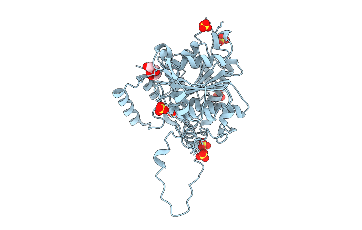
Crystal structure of PhaC1 from Ralstonia eutropha
Biological Source:
Source Organism:
Host Organism:
Method Details:
Experimental Method:
Resolution:
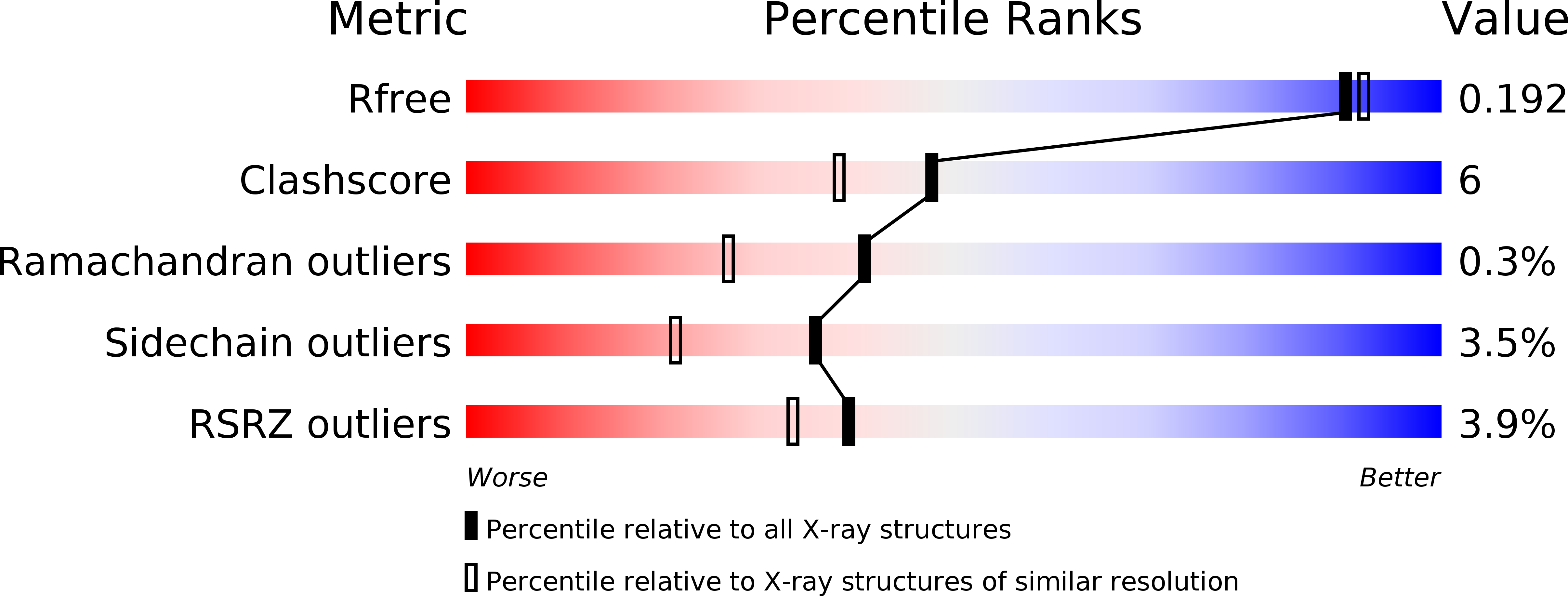
1.80 Å
R-Value Free:
0.18
R-Value Work:
0.15
R-Value Observed:
0.15
Space Group:
I 2 2 2
Macromolecular Entities
Polymer Type:polypeptide(L)
Molecule:Poly-beta-hydroxybutyrate polymerase
Gene (Uniprot):phaC
Chain IDs:A
Chain Length:396
Number of Molecules:1
Biological Source:Cupriavidus necator
Primary Citation
Crystal structure of Ralstonia eutropha polyhydroxyalkanoate synthase C-terminal domain and reaction mechanisms.
Biotechnol J 12 ? ? (2017)
PMID: 27808482 DOI: 10.1002/biot.201600648

Abstact

Polyhydroxyalkanoates (PHAs) are natural polyesters synthesized by numerous microorganisms as energy and reducing power storage materials, and have attracted much attention as substitutes for petroleum-based plastics. Here, we report the first crystal structure of Ralstonia eutropha PHA synthase at 1.8 Å resolution and structure-based mechanisms for PHA polymerization. RePhaC1 contains two distinct domains, the N-terminal (RePhaC1ND) and C-terminal domains (RePhaC1CD), and exists as a dimer. RePhaC1CD catalyzes polymerization via non-processive ping-pong mechanism using a Cys-His-Asp catalytic triad. Molecular docking simulation of 3-hydroxybutyryl-CoA to the active site of RePhaC1CD reveals residues involved in the formation of 3-hydroxybutyryl-CoA binding pocket and substrate binding tunnel. Comparative analysis with other polymerases elucidates how different classes of PHA synthases show different substrate specificities. Furthermore, we attempted structure-based protein engineering and developed a RePhaC1 mutant with enhanced PHA synthase activity.

Legend

Protein

Chemical

Disease

Primary Citation of related structures