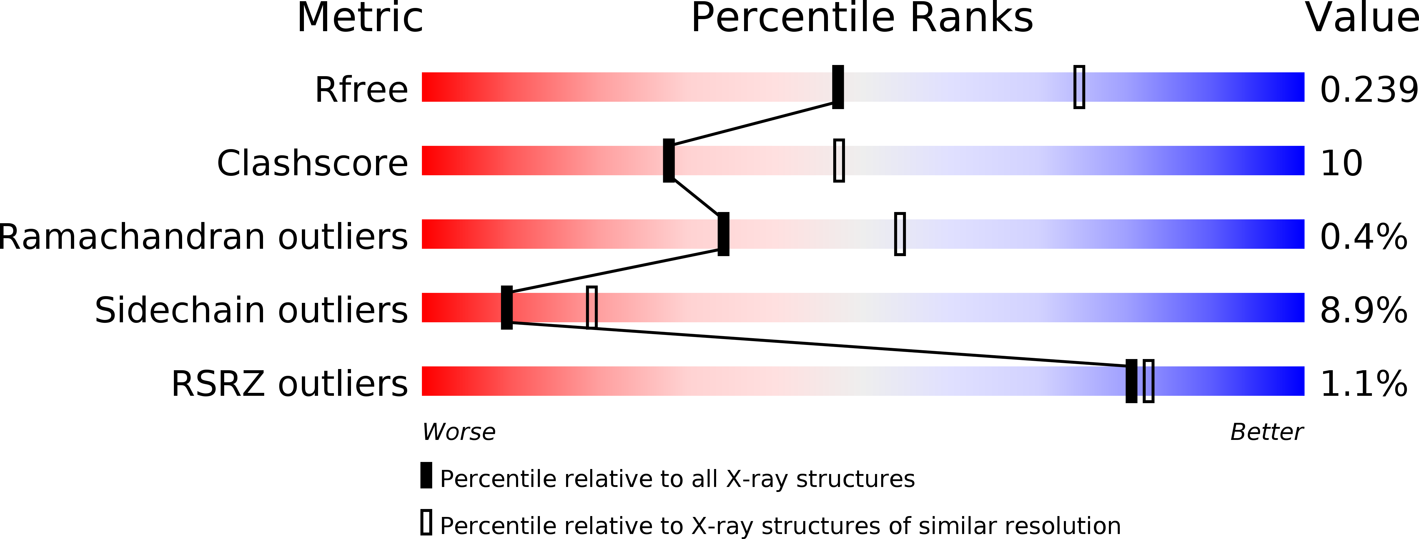
5HXY image
Deposition Date 2016-01-31
Release Date 2017-02-01
Last Version Date 2024-11-20
Entry Detail
PDB ID:
5HXY
Keywords:
Title:
Crystal structure of XerA recombinase
Biological Source:
Source Organism:
Method Details:
Experimental Method:
Resolution:
2.50 Å
R-Value Free:
0.24
R-Value Work:
0.18
R-Value Observed:
0.19
Space Group:
C 1 2 1
Macromolecular Entities
Polymer Type:polypeptide(L)
Molecule:Tyrosine recombinase XerA
Gene (Uniprot):xerA
Chain IDs:A, B, C, D, E, F
Chain Length:317
Number of Molecules:6
Biological Source:Thermoplasma acidophilum DSM 1728
Modified Residue
Compound ID Chain ID Parent Comp ID Details 2D Image
MSE A MET modified residue
Ligand Molecules
Primary Citation
Crystal structure of Thermoplasma acidophilum XerA recombinase shows large C-shape clamp conformation and cis-cleavage mode for nucleophilic tyrosine
FEBS Lett. 590 848 856 (2016)
PMID: 26919387 DOI: 10.1002/1873-3468.12109

Abstact

Site-specific Xer recombination plays a pivotal role in reshuffling genetic information. Here, we report the 2.5 Å crystal structure of XerA from the archaean Thermoplasma acidophilum. Crystallographic data reveal a uniquely open conformational state, resulting in a C-shaped clamp with an angle of ~ 48° and a distance of 57 Å between the core-binding and the catalytic domains. The catalytic nucleophile, Tyr264, is positioned in cis-cleavage mode by XerA's C-term tail that interacts with the CAT domain of a neighboring monomer without DNA substrate. Structural comparisons of tyrosine recombinases elucidate the dynamics of Xer recombinase.

Legend

Protein

Chemical

Disease

Primary Citation of related structures