
Deposition Date
2016-01-29
Release Date
2016-05-18
Last Version Date
2023-11-08
Entry Detail
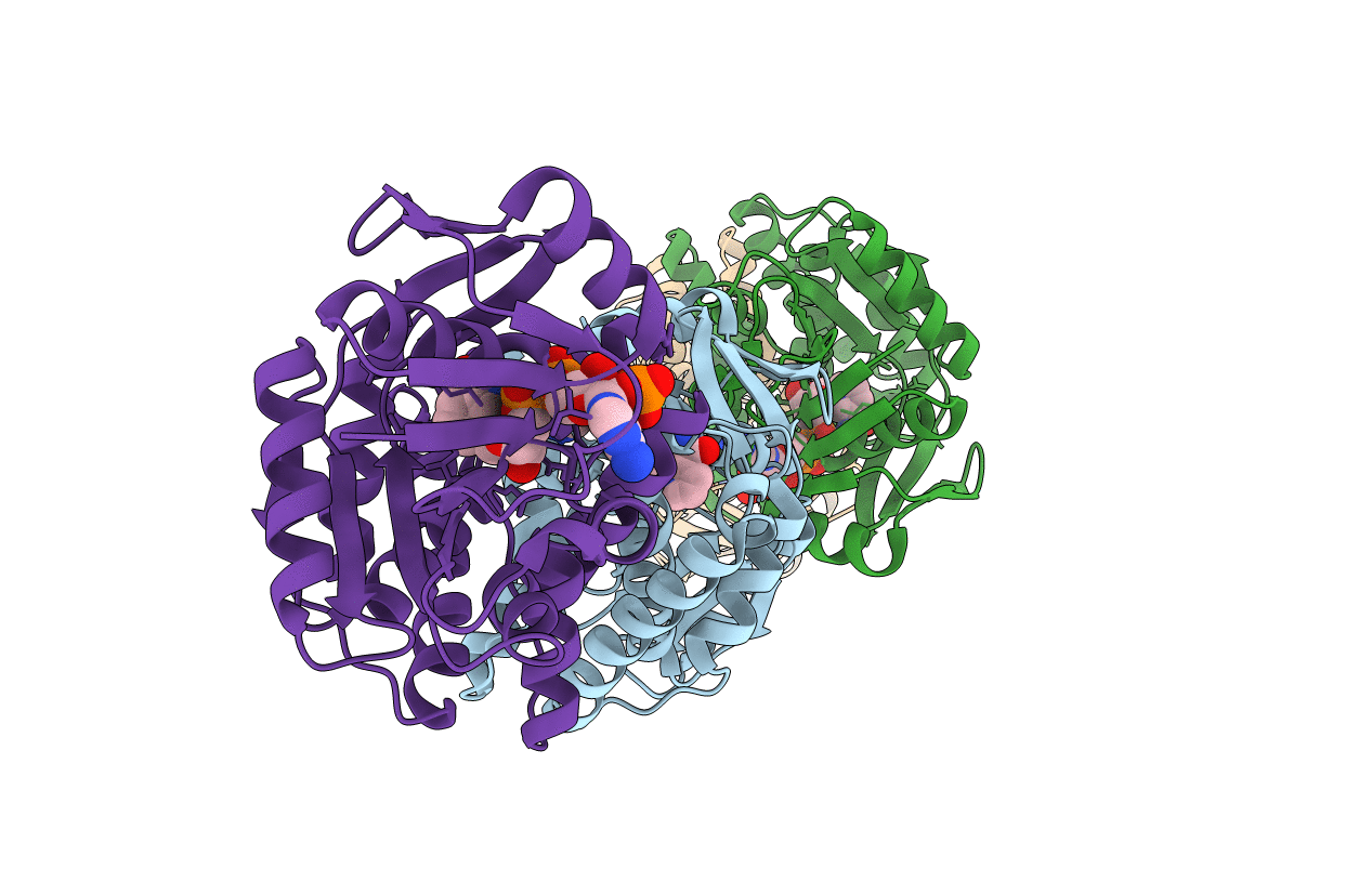
PDB ID:
5HWS
Keywords:
Title:
Crystal structure of ketopantoate reductase from Thermococcus kodakarensis complexed with NADP+
Biological Source:
Source Organism(s):
Expression System(s):
Method Details:
Experimental Method:
Resolution:
2.30 Å
R-Value Free:
0.26
R-Value Work:
0.22
R-Value Observed:
0.22
Space Group:
P 1
