Deposition Date
2016-01-28
Release Date
2016-04-20
Last Version Date
2024-11-06
Entry Detail
PDB ID:
5HVZ
Keywords:
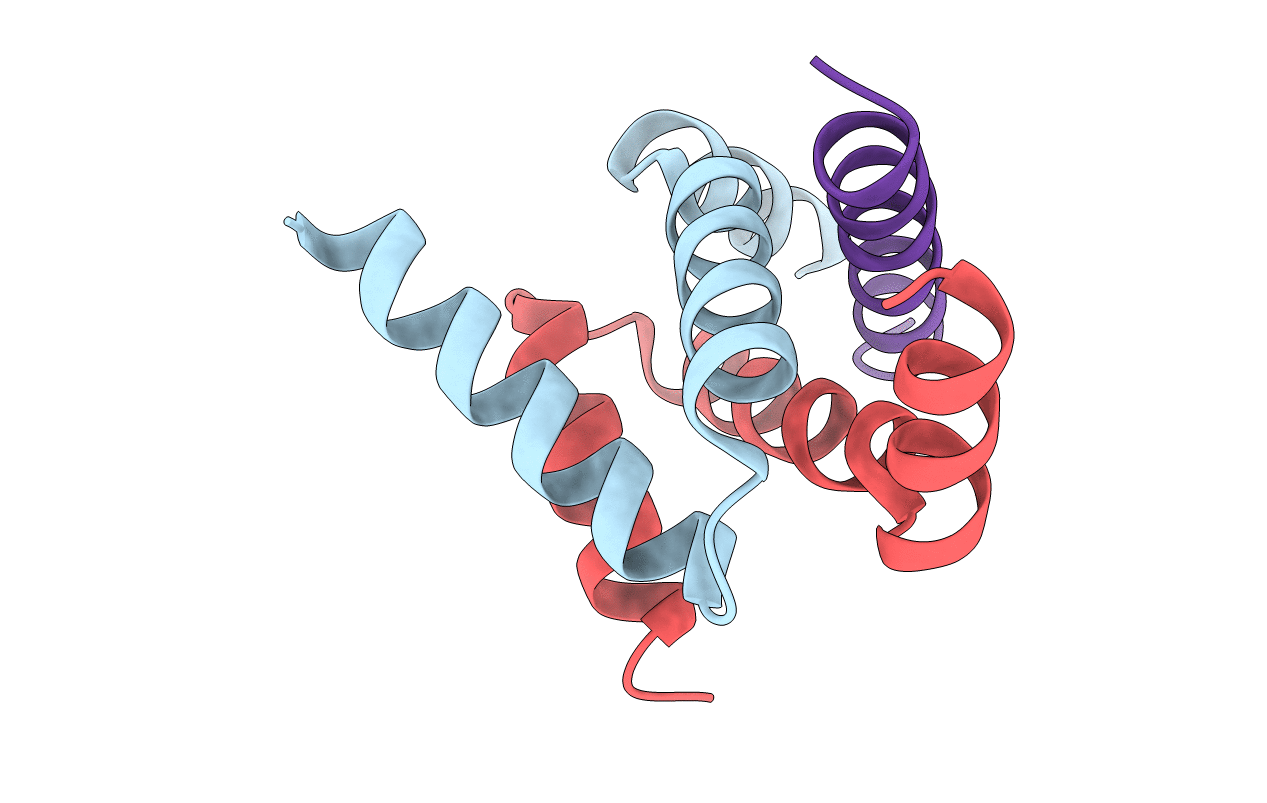
Title:
Crystal structure of smAKAP AKB domain bound RIa dimerization/docking (D/D) complex at 2.0 A resolution
Biological Source:
Source Organism(s):
Bos taurus (Taxon ID: 9913)
Homo sapiens (Taxon ID: 9606)
Homo sapiens (Taxon ID: 9606)
Expression System(s):
Method Details:
Experimental Method:
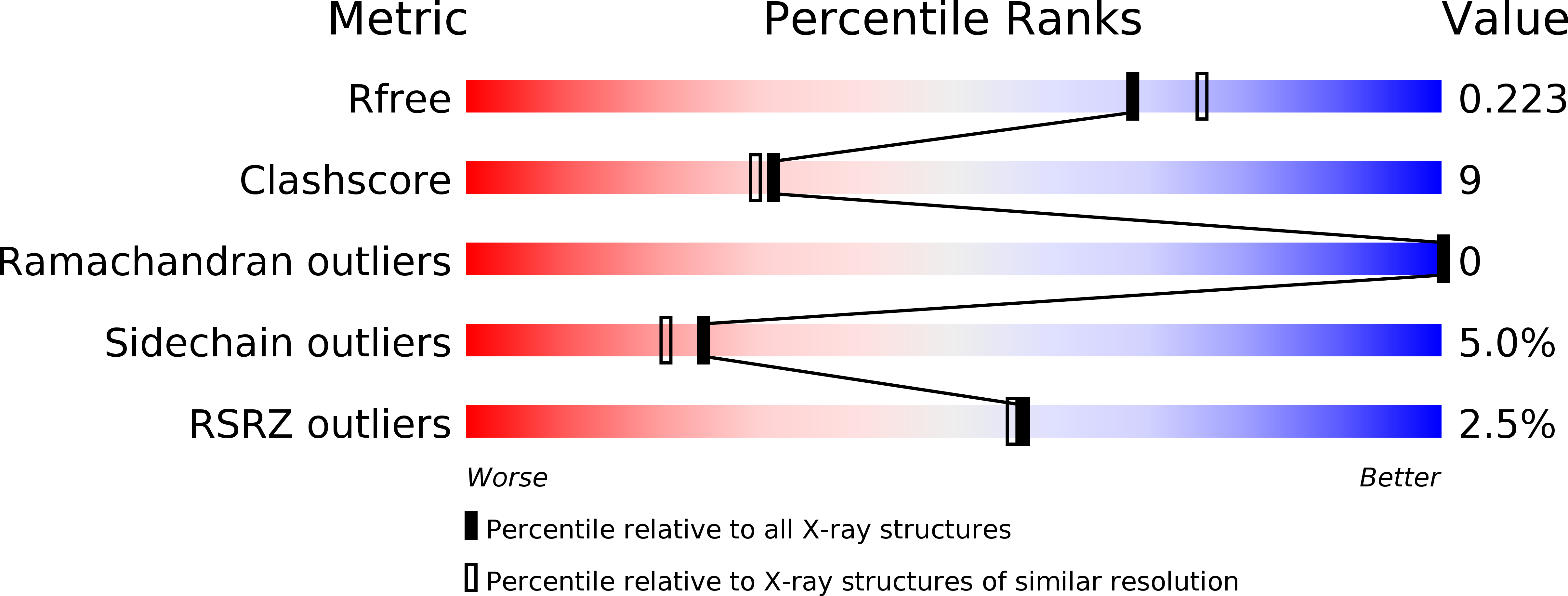
Resolution:
2.00 Å
R-Value Free:
0.24
R-Value Work:
0.20
R-Value Observed:
0.21
Space Group:
P 21 21 21