
Deposition Date
2016-01-28
Release Date
2016-03-09
Last Version Date
2024-06-19
Entry Detail
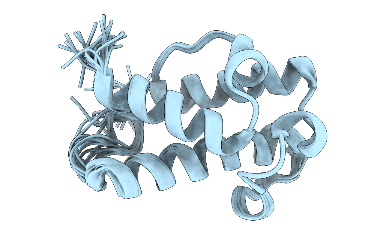
PDB ID:
5HVC
Keywords:
Title:
Solution structure of the apo state of the acyl carrier protein from the MLSA2 subunit of the mycolactone polyketide synthase
Biological Source:
Source Organism(s):
Mycobacterium ulcerans (Taxon ID: 1809)
Expression System(s):
Method Details:
Experimental Method:
Conformers Calculated:
50
Conformers Submitted:
20
Selection Criteria:
structures with the lowest energy
