5HUZ image
Deposition Date 2016-01-27
Release Date 2016-03-02
Last Version Date 2024-05-15
Entry Detail
PDB ID:
5HUZ
Title:
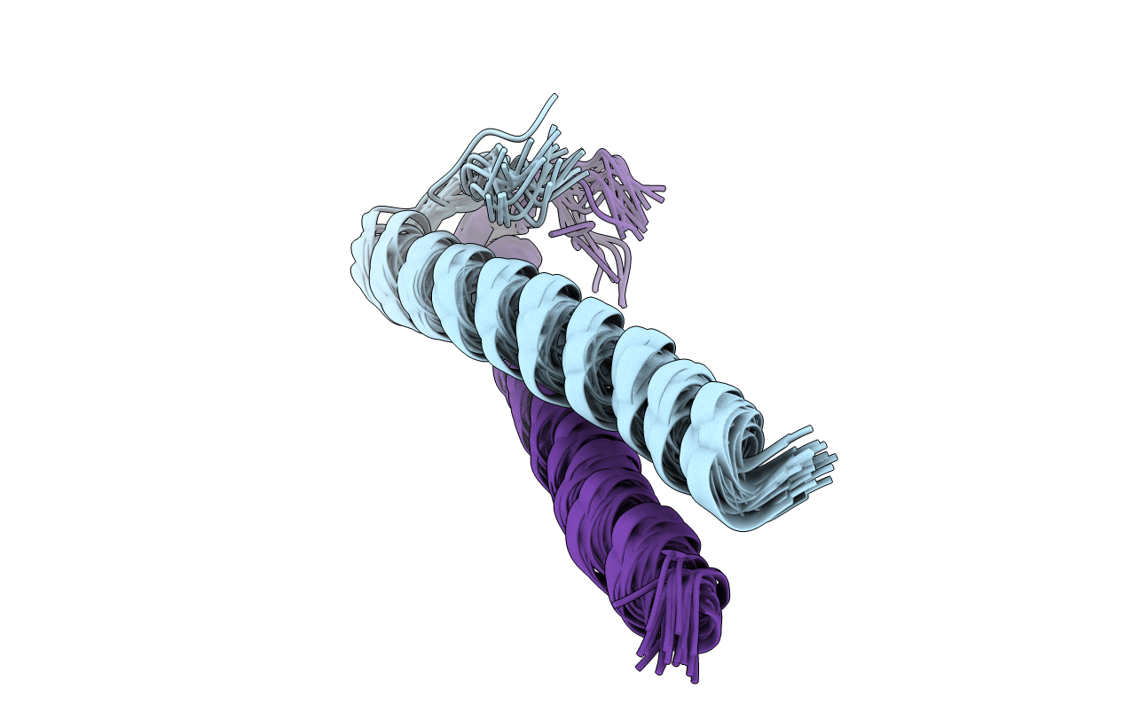
Solution structure of coiled coil domain of myosin binding subunit of myosin light chain phosphatase
Biological Source:
Source Organism:
Homo sapiens (Taxon ID: 9606)
Host Organism:
Method Details:
Experimental Method:
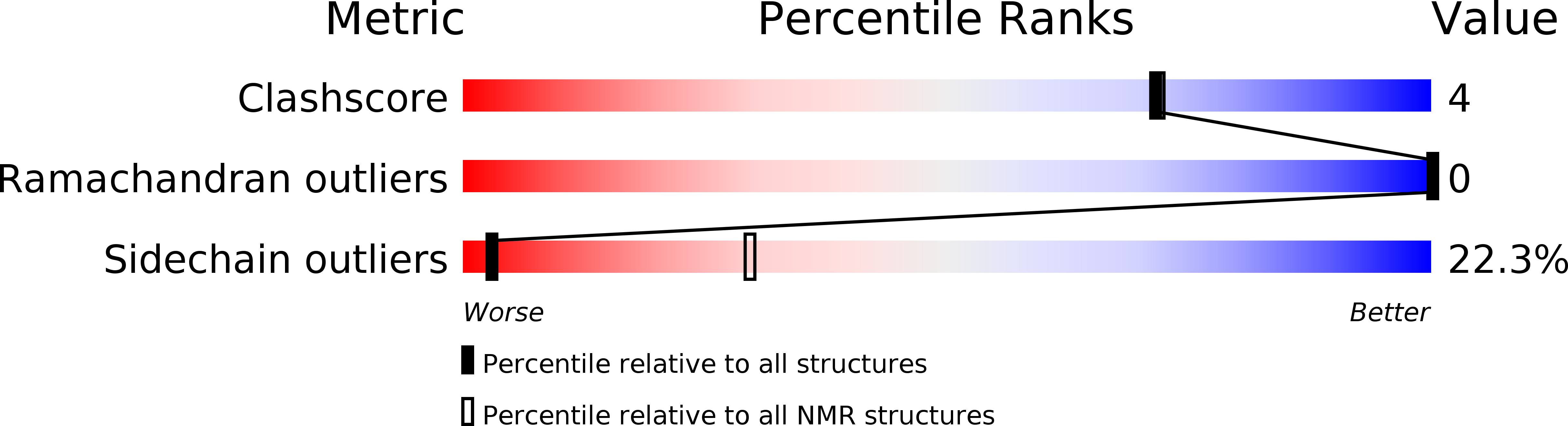
Conformers Calculated:
20
Conformers Submitted:
20
Selection Criteria:
structures with the least restraint violations
Macromolecular Entities
Structures with similar UniProt ID
Protein Blast
Polymer Type:polypeptide(L)
Molecule:Protein phosphatase 1 regulatory subunit 12A
Gene (Uniprot):PPP1R12A
Chain IDs:A, B
Chain Length:48
Number of Molecules:2
Biological Source:Homo sapiens
Ligand Molecules
Primary Citation
NMR assignment and secondary structure of coiled coil domain of C-terminal myosin binding subunit of myosin phosphatase
Protein Pept Lett. 21(7) 639 645 (2014)
PMID: 24693955

Abstact

Protein-protein interactions between the C-terminal domain of Myosin Binding Subunit (MBS) of MLC Phosphatase (MBS(CT180); C-terminal 180 aa) and the N-terminal coiled coil (CC) leucine zipper (LZ) domain of PKGIα, PKG-Iα(1-159) play an important role in the process of Smooth Muscle Cell relaxation. The paucity of three-dimensional structural information for MBS(CT180) prevents an atomic level understanding of the MBS-PKG contractile complex. MBS(CT180) is comprised of three structurally different sub-domains including a non-canonical CC, a CC, and a LZ. Recently we reported polypeptide purification and biophysical characterization of the CC domain and the LZ domain of MBS(CT180) (Sharma et al, Prot Expr Purif 2012). Here we report (1)H, (13)C, (15)N chemical shift assignments of homodimeric CC MBS domain encompassing amino acid residues Asp931-Leu980 using 2D and 3D heteronuclear NMR spectroscopy. Secondary structure analyses deduced from these NMR chemical shift data have identified a contiguous stretch of 36 residues from Phe932 to Ala967 that is involved in the formation of coiled coil α-helical region within CC MBS domain. The N-terminal residue Asp931 and the C-terminally positioned residues Thr968-Ala975, Arg977, and Ser978 adopt nonhelical loop conformations.

Legend

Protein

Chemical

Disease

Primary Citation of related structures
Feedback Form
Name
Email
Institute
Feedback