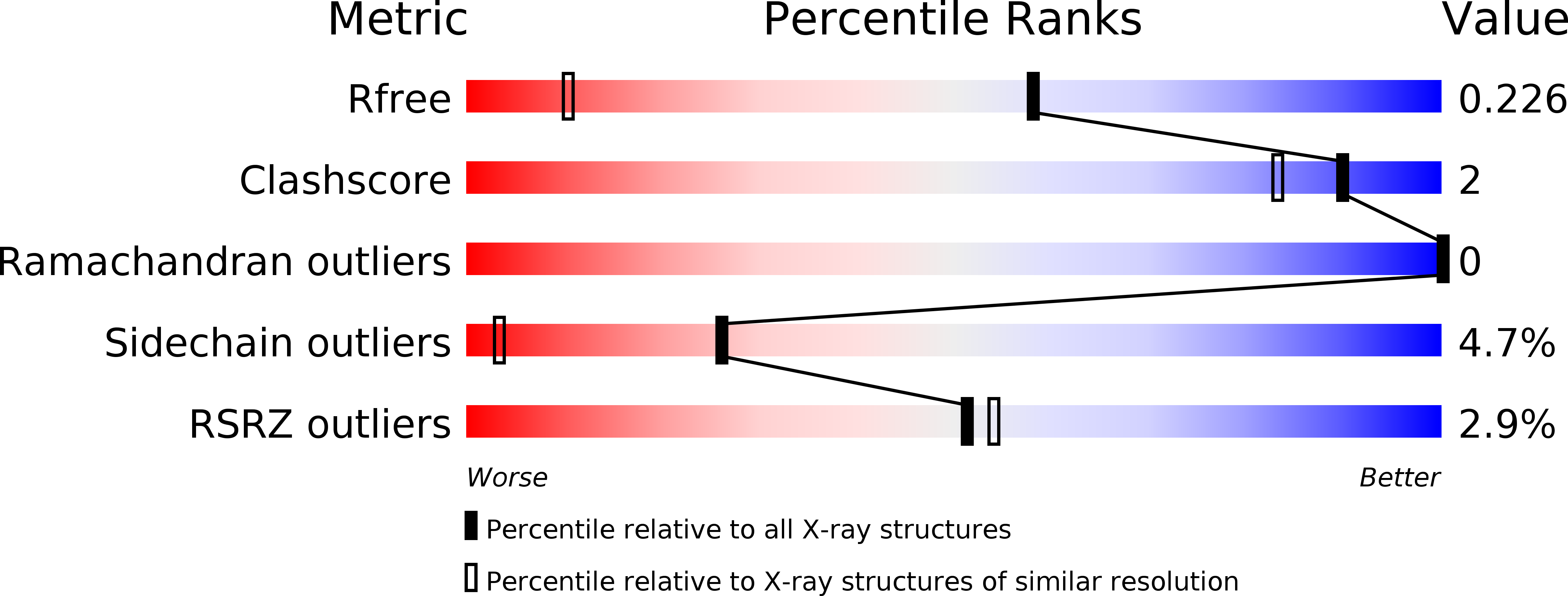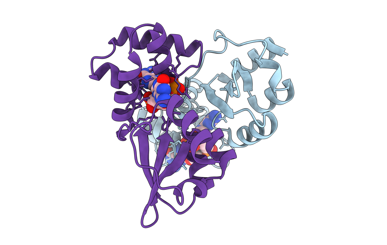
5HTL image
Deposition Date 2016-01-27
Release Date 2016-10-05
Last Version Date 2024-10-09
Entry Detail
PDB ID:
5HTL
Title:
Structure of MshE with cdg
Biological Source:
Source Organism:
Vibrio cholerae (Taxon ID: 666)
Method Details:
Experimental Method:
Resolution:
1.37 Å
R-Value Free:
0.22
R-Value Work:
0.20
R-Value Observed:
0.20
Space Group:
P 21 21 21
Macromolecular Entities
Polymer Type:polypeptide(L)
Molecule:MSHA biogenesis protein MshE
Chain IDs:A, B
Chain Length:145
Number of Molecules:2
Biological Source:Vibrio cholerae
Modified Residue
Compound ID Chain ID Parent Comp ID Details 2D Image
MSE A MET modified residue
Ligand Molecules
Primary Citation
Nucleotide binding by the widespread high-affinity cyclic di-GMP receptor MshEN domain.
Nat Commun 7 12481 12481 (2016)
PMID: 27578558 DOI: 10.1038/ncomms12481

Abstact

C-di-GMP is a bacterial second messenger regulating various cellular functions. Many bacteria contain c-di-GMP-metabolizing enzymes but lack known c-di-GMP receptors. Recently, two MshE-type ATPases associated with bacterial type II secretion system and type IV pilus formation were shown to specifically bind c-di-GMP. Here we report crystal structure of the MshE N-terminal domain (MshEN1-145) from Vibrio cholerae in complex with c-di-GMP at a 1.37 Å resolution. This structure reveals a unique c-di-GMP-binding mode, featuring a tandem array of two highly conserved binding motifs, each comprising a 24-residue sequence RLGxx(L/V/I)(L/V/I)xxG(L/V/I)(L/V/I)xxxxLxxxLxxQ that binds half of the c-di-GMP molecule, primarily through hydrophobic interactions. Mutating these highly conserved residues markedly reduces c-di-GMP binding and biofilm formation by V. cholerae. This c-di-GMP-binding motif is present in diverse bacterial proteins exhibiting binding affinities ranging from 0.5 μM to as low as 14 nM. The MshEN domain contains the longest nucleotide-binding motif reported to date.

Legend

Protein

Chemical

Disease

Primary Citation of related structures