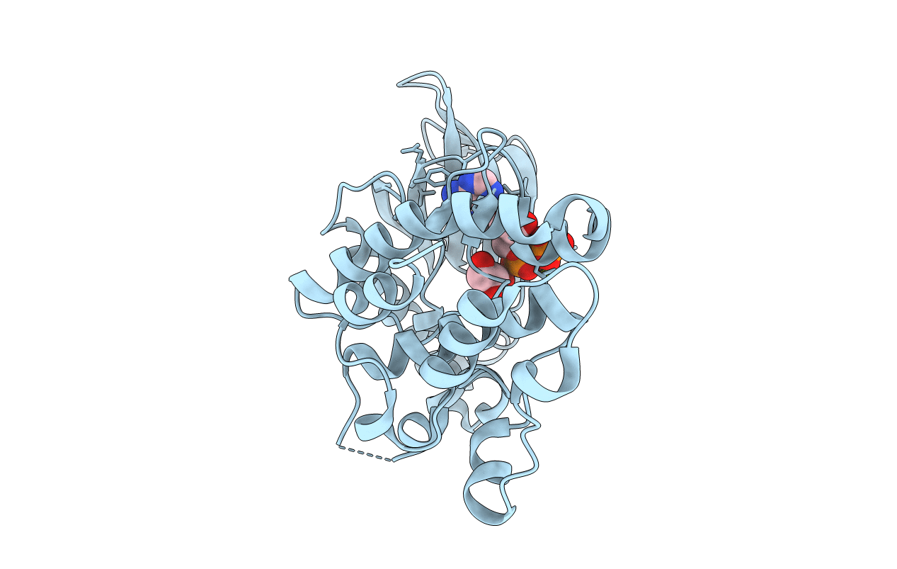
5HNV image
Entry Detail
PDB ID:
5HNV
Keywords:
Title:
Crystal structure of PpkA
Biological Source:
Source Organism:
Host Organism:
PDB Version:
Deposition Date:
2016-01-19
Release Date:
2017-01-25
Method Details:
Experimental Method:
Resolution:
1.41 Å
R-Value Free:
0.21
R-Value Work:
0.18
R-Value Observed:
0.18
Space Group:
P 21 21 2
Macromolecular Entities
Polymer Type:polypeptide(L)
Description:PpkA N terminal
Chain IDs:A
Chain Length:302
Number of Molecules:1
Biological Source:Serratia sp. FS14
Primary Citation
Crystal structures of the kinase domain of PpkA, a key regulatory component of T6SS, reveal a general inhibitory mechanism.
Biochem.J. 475 2209 2224 (2018)
PMID: 29858276 DOI: 10.1042/BCJ20180077

Abstact

The type VI secretion system (T6SS) is a versatile and widespread export system found in many Gram-negative bacteria that delivers effector proteins into target cells. The functions of T6SSs are tightly regulated by diverse mechanisms at multiple levels, including post-translational modification through threonine phosphorylation via the Ser/Thr protein kinase (STPK) PpkA. Here, we identified that PpkA is essential for T6SS secretion in Serratia marcescens since its deletion eliminated the secretion of haemolysin co-regulated protein, while the periplasmic and transmembrane portion of PpkA was found to be disposable for T6SS secretion. We further determined the crystal structure of the kinase domain of PpkA (PpkA-294). The structure of PpkA-294 was determined in its apo form to a 1.6 Å resolution as well as in complex with ATP to a 1.41 Å resolution and with an ATP analogue AMP-PCP to a 1.45 Å resolution. The residues in the activation loop of PpkA-294 were fully determined, and the N-terminus of the loop was folded into an unprecedented inhibitory helix, revealing that the PpkA kinase domain was in an auto-inhibitory state. The ternary MgATP-PpkA-294 complex was also inactive with nucleotide ribose and phosphates in unexpected and unproductive conformations. The αC-helix in the inactive PpkA-294 adopted a conformation towards the active site but with the conserved glutamate in the helix rotated away, which we suggest to be a general conformation for all STPK kinases in the inactive form. Structural comparison of PpkA with its eukaryotic homologues reinforced the universal regulation mechanism of protein kinases.

Legend

Protein

Chemical

Disease

Primary Citation of related structures