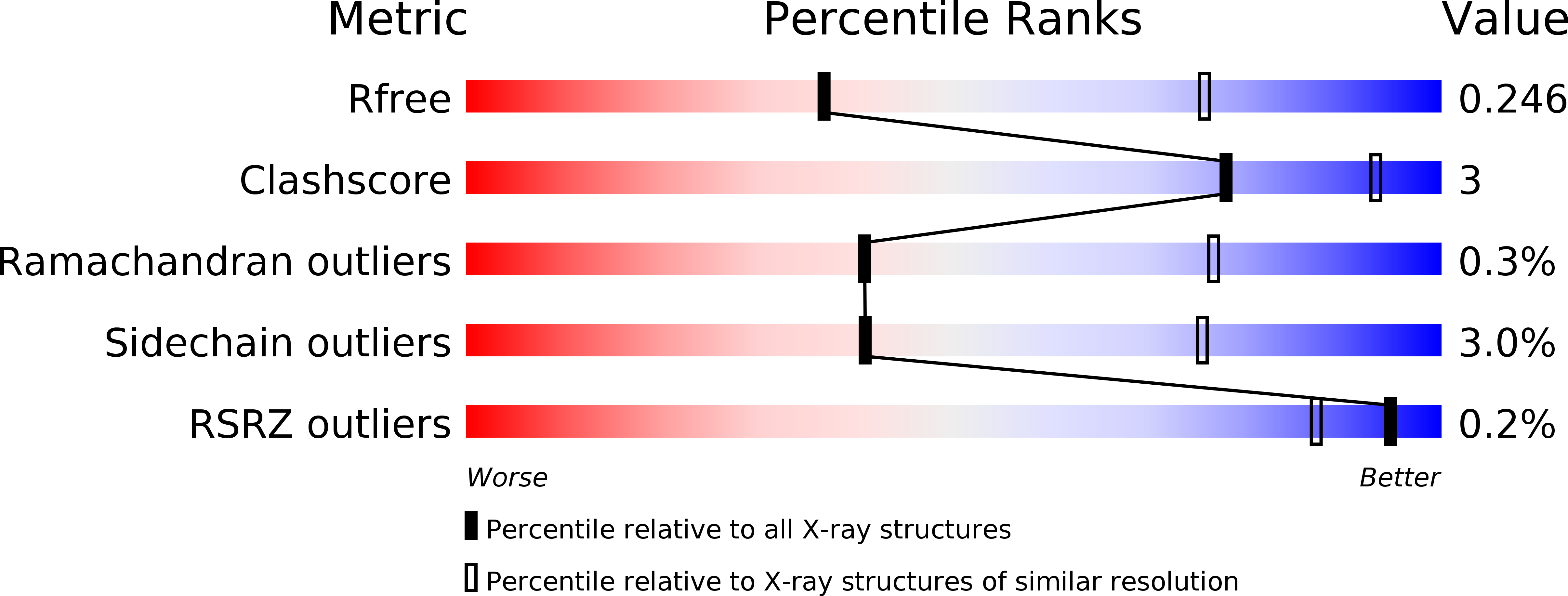
5HKK image
Deposition Date 2016-01-14
Release Date 2016-09-21
Last Version Date 2024-01-10
Entry Detail
PDB ID:
5HKK
Keywords:
Title:
Caldalaklibacillus thermarum F1-ATPase (wild type)
Biological Source:
Source Organism:
Method Details:
Experimental Method:
Resolution:
3.00 Å
R-Value Free:
0.24
R-Value Work:
0.20
R-Value Observed:
0.20
Space Group:
P 1 21 1
Macromolecular Entities
Polymer Type:polypeptide(L)
Molecule:ATP synthase subunit alpha
Gene (Uniprot):atpA
Chain IDs:A, B, C, I, J, K
Chain Length:502
Number of Molecules:6
Biological Source:Caldalkalibacillus thermarum TA2.A1
Polymer Type:polypeptide(L)
Molecule:ATP synthase subunit beta
Gene (Uniprot):atpD
Chain IDs:D, E, F, L, M, N
Chain Length:462
Number of Molecules:6
Biological Source:Caldalkalibacillus thermarum TA2.A1
Polymer Type:polypeptide(L)
Molecule:ATP synthase gamma chain
Gene (Uniprot):atpG
Chain IDs:G, O
Chain Length:286
Number of Molecules:2
Biological Source:Caldalkalibacillus thermarum TA2.A1
Polymer Type:polypeptide(L)
Molecule:ATP synthase epsilon chain
Gene (Uniprot):atpC
Chain IDs:H, P
Chain Length:135
Number of Molecules:2
Biological Source:Caldalkalibacillus thermarum TA2.A1
Primary Citation
Regulation of the thermoalkaliphilic F1-ATPase from Caldalkalibacillus thermarum.
Proc.Natl.Acad.Sci.USA 113 10860 10865 (2016)
PMID: 27621435 DOI: 10.1073/pnas.1612035113

Abstact

The crystal structure has been determined of the F1-catalytic domain of the F-ATPase from Caldalkalibacillus thermarum, which hydrolyzes adenosine triphosphate (ATP) poorly. It is very similar to those of active mitochondrial and bacterial F1-ATPases. In the F-ATPase from Geobacillus stearothermophilus, conformational changes in the ε-subunit are influenced by intracellular ATP concentration and membrane potential. When ATP is plentiful, the ε-subunit assumes a "down" state, with an ATP molecule bound to its two C-terminal α-helices; when ATP is scarce, the α-helices are proposed to inhibit ATP hydrolysis by assuming an "up" state, where the α-helices, devoid of ATP, enter the α3β3-catalytic region. However, in the Escherichia coli enzyme, there is no evidence that such ATP binding to the ε-subunit is mechanistically important for modulating the enzyme's hydrolytic activity. In the structure of the F1-ATPase from C. thermarum, ATP and a magnesium ion are bound to the α-helices in the down state. In a form with a mutated ε-subunit unable to bind ATP, the enzyme remains inactive and the ε-subunit is down. Therefore, neither the γ-subunit nor the regulatory ATP bound to the ε-subunit is involved in the inhibitory mechanism of this particular enzyme. The structure of the α3β3-catalytic domain is likewise closely similar to those of active F1-ATPases. However, although the βE-catalytic site is in the usual "open" conformation, it is occupied by the unique combination of an ADP molecule with no magnesium ion and a phosphate ion. These bound hydrolytic products are likely to be the basis of inhibition of ATP hydrolysis.

Legend

Protein

Chemical

Disease

Primary Citation of related structures