5HJ3 image
Deposition Date 2016-01-12
Release Date 2016-03-09
Last Version Date 2024-10-30
Entry Detail
PDB ID:
5HJ3
Title:
Crystal structure of host-primed Ebola virus GP, GPcl.
Biological Source:
Source Organism:
Host Organism:
Method Details:
Experimental Method:
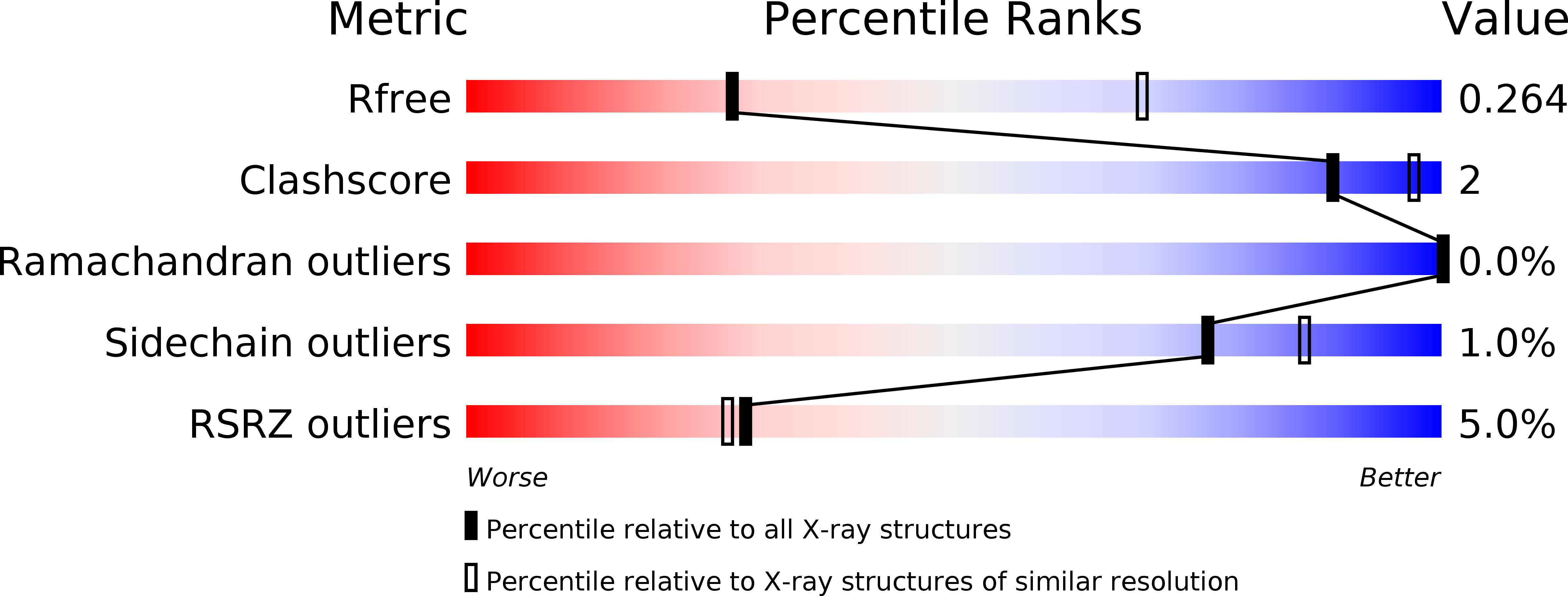
Resolution:
3.30 Å
R-Value Free:
0.25
R-Value Work:
0.22
R-Value Observed:
0.22
Space Group:
H 3
Macromolecular Entities
Polymer Type:polypeptide(L)
Molecule:Envelope glycoprotein
Gene (Uniprot):GP
Chain IDs:A (auth: C), E (auth: G), I (auth: K), M (auth: O)
Chain Length:179
Number of Molecules:4
Biological Source:Ebola virus sp.
Polymer Type:polypeptide(L)
Molecule:Envelope glycoprotein
Gene (Uniprot):GP
Chain IDs:B (auth: D), F (auth: H), J (auth: L), N (auth: P)
Chain Length:136
Number of Molecules:4
Biological Source:Zaire ebolavirus
Polymer Type:polypeptide(L)
Molecule:KZ52 Antibody Fragment
Chain IDs:C (auth: E), G (auth: I), K (auth: M), O (auth: A)
Chain Length:226
Number of Molecules:4
Biological Source:Homo sapiens
Polymer Type:polypeptide(L)
Molecule:KZ52 Antibody Fragment
Chain IDs:D (auth: F), H (auth: J), L (auth: N), P (auth: B)
Chain Length:217
Number of Molecules:4
Biological Source:Homo sapiens
Ligand Molecules
Primary Citation
Host-Primed Ebola Virus GP Exposes a Hydrophobic NPC1 Receptor-Binding Pocket, Revealing a Target for Broadly Neutralizing Antibodies.
Mbio 7 e02154 e02115 (2016)
PMID: 26908579 DOI: 10.1128/mBio.02154-15

Abstact

UNLABELLED The filovirus surface glycoprotein (GP) mediates viral entry into host cells. Following viral internalization into endosomes, GP is cleaved by host cysteine proteases to expose a receptor-binding site (RBS) that is otherwise hidden from immune surveillance. Here, we present the crystal structure of proteolytically cleaved Ebola virus GP to a resolution of 3.3 Å. We use this structure in conjunction with functional analysis of a large panel of pseudotyped viruses bearing mutant GP proteins to map the Ebola virus GP endosomal RBS at molecular resolution. Our studies indicate that binding of GP to its endosomal receptor Niemann-Pick C1 occurs in two distinct stages: the initial electrostatic interactions are followed by specific interactions with a hydrophobic trough that is exposed on the endosomally cleaved GP1 subunit. Finally, we demonstrate that monoclonal antibodies targeting the filovirus RBS neutralize all known filovirus GPs, making this conserved pocket a promising target for the development of panfilovirus therapeutics. IMPORTANCE Ebola virus uses its glycoprotein (GP) to enter new host cells. During entry, GP must be cleaved by human enzymes in order for receptor binding to occur. Here, we provide the crystal structure of the cleaved form of Ebola virus GP. We demonstrate that cleavage exposes a site at the top of GP and that this site binds the critical domain C of the receptor, termed Niemann-Pick C1 (NPC1). We perform mutagenesis to find parts of the site essential for binding NPC1 and map distinct roles for an upper, charged crest and lower, hydrophobic trough in cleaved GP. We find that this 3-dimensional site is conserved across the filovirus family and that antibody directed against this site is able to bind cleaved GP from every filovirus tested and neutralize viruses bearing those GPs.

Legend

Protein

Chemical

Disease

Primary Citation of related structures