Deposition Date
2016-01-08
Release Date
2017-04-12
Last Version Date
2024-03-06
Entry Detail
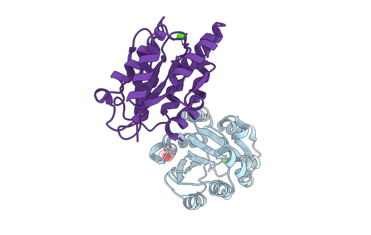
PDB ID:
5HGJ
Keywords:
Title:
Structure of integrin alpha1beta1 and alpha2beta1 I-domains explain differential calcium-mediated ligand recognition
Biological Source:
Source Organism(s):
Homo sapiens (Taxon ID: 9606)
Expression System(s):
Method Details:
Experimental Method:
Resolution:
1.40 Å
R-Value Free:
0.20
R-Value Work:
0.17
R-Value Observed:
0.17
Space Group:
P 1 21 1