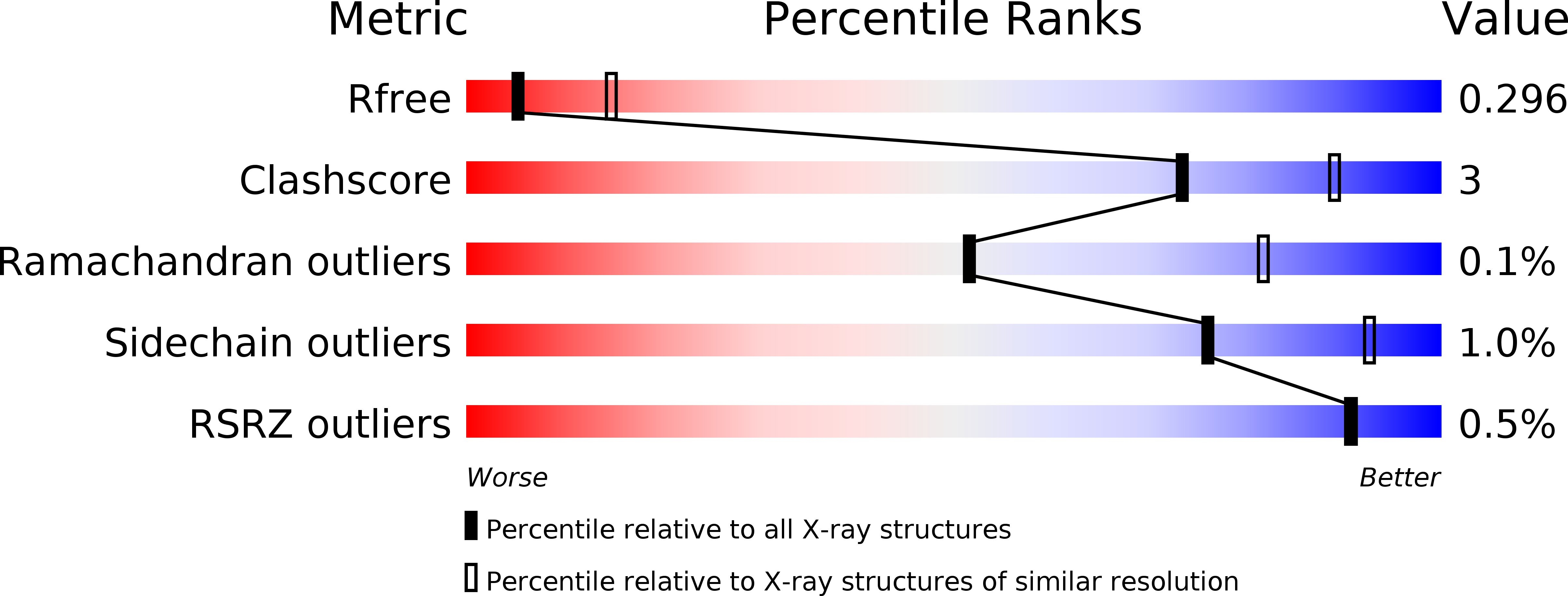
5HDP image
Deposition Date 2016-01-05
Release Date 2017-01-11
Last Version Date 2024-11-20
Entry Detail
PDB ID:
5HDP
Keywords:
Title:
Hydrolase StnA mutant - S185A
Biological Source:
Source Organism:
Method Details:
Experimental Method:
Resolution:
2.90 Å
R-Value Free:
0.29
R-Value Work:
0.24
R-Value Observed:
0.24
Space Group:
P 1
Macromolecular Entities
Structures with similar UniProt ID
Protein Blast
Polymer Type:polypeptide(L)
Molecule:Hydrolase
Gene (Uniprot):stnA
Mutagens:S185A
Chain IDs:A (auth: D), B (auth: A), C (auth: B), D (auth: C), E, F, G
Chain Length:383
Number of Molecules:7
Biological Source:Streptomyces flocculus
Ligand Molecules
Primary Citation
Crystal Structure of StnA for the Biosynthesis of Antitumor Drug Streptonigrin Reveals a Unique Substrate Binding Mode
Sci Rep 7 40254 40254 (2017)
PMID: 28074848 DOI: 10.1038/srep40254

Abstact

Streptonigrin methylesterase A (StnA) is one of the tailoring enzymes that modify the aminoquinone skeleton in the biosynthesis pathway of Streptomyces species. Although StnA has no significant sequence homology with the reported α/β-fold hydrolases, it shows typical hydrolytic activity in vivo and in vitro. In order to reveal its functional characteristics, the crystal structures of the selenomethionine substituted StnA (SeMet-StnA) and the complex (S185A mutant) with its substrate were resolved to the resolution of 2.71 Å and 2.90 Å, respectively. The overall structure of StnA can be described as an α-helix cap domain on top of a common α/β hydrolase domain. The substrate methyl ester of 10'-demethoxystreptonigrin binds in a hydrophobic pocket that mainly consists of cap domain residues and is close to the catalytic triad Ser185-His349-Asp308. The transition state is stabilized by an oxyanion hole formed by the backbone amides of Ala102 and Leu186. The substrate binding appears to be dominated by interactions with several specific hydrophobic contacts and hydrogen bonds in the cap domain. The molecular dynamics simulation and site-directed mutagenesis confirmed the important roles of the key interacting residues in the cap domain. Structural alignment and phylogenetic tree analysis indicate that StnA represents a new subfamily of lipolytic enzymes with the specific binding pocket located at the cap domain instead of the interface between the two domains.

Legend

Protein

Chemical

Disease

Primary Citation of related structures