5HBC image
Deposition Date 2015-12-31
Release Date 2016-01-20
Last Version Date 2024-10-16
Entry Detail
PDB ID:
5HBC
Keywords:
Title:
Intermediate structure of iron-saturated C-lobe of bovine lactoferrin at 2.79 Angstrom resolution indicates the softening of iron coordination
Biological Source:
Source Organism:
Bos taurus (Taxon ID: 9913)
Method Details:
Experimental Method:
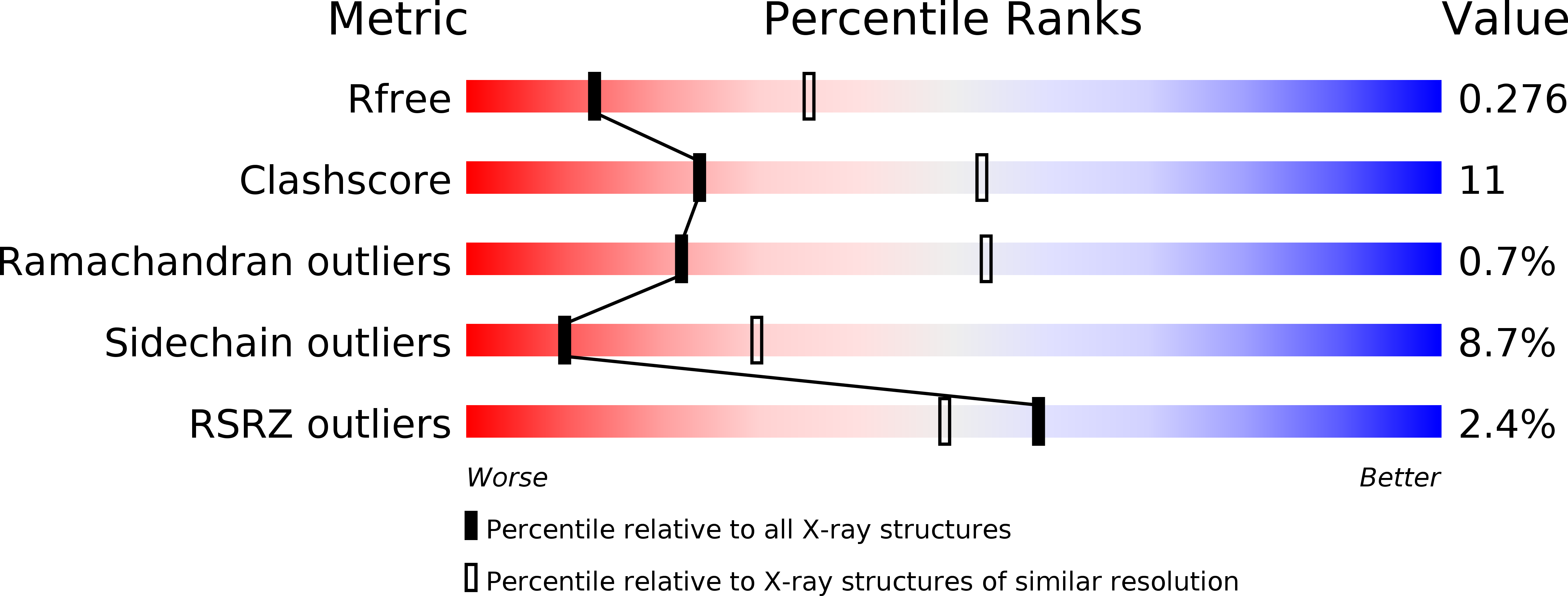
Resolution:
2.79 Å
R-Value Free:
0.27
R-Value Work:
0.20
R-Value Observed:
0.20
Space Group:
C 1 2 1
Macromolecular Entities
Structures with similar UniProt ID
Protein Blast
Polymer Type:polypeptide(L)
Molecule:Lactotransferrin
Gene (Uniprot):LTF
Chain IDs:A, B
Chain Length:348
Number of Molecules:2
Biological Source:Bos taurus
Primary Citation
Structure of iron saturated C-lobe of bovine lactoferrin at pH 6.8 indicates a weakening of iron coordination
Proteins 84 591 599 (2016)
PMID: 26850578 DOI: 10.1002/prot.25004

Abstact

The bilobal lactoferrin is an approximately 76 kDa glycoprotein. It sequesters two Fe(3+) ions together with two CO(3)(2-) ions. The C-terminal half (residues, Tyr342-Arg689, C-lobe) of bovine lactoferrin (BLF) (residues Ala1-Arg689) was prepared by limited proteolysis using trypsin. Both C-lobe and intact BLF were saturated to 100%. Both of them retained up to nearly 85% of iron at pH 6.5. At pH 5.0, C-lobe retained 75% of iron whereas intact protein could retain only slightly more than 60%. At pH 4.0 both contained 25% iron and at pH 2.0 they were left with iron concentration of only 10%. The structure of iron saturated C-lobe was determined at 2.79 Å resolution and refined to R(cryst) and R(free) factors of 0.205 and 0.273, respectively. The structure contains two crystallographically independent molecules, A and B. They were found to have identical structures with an r.m.s. shift of 0.5 Å for their C(α) atoms. A high solvent content of 66% was observed in the crystals. The average value of an overall B-factor was 68.0 Å(2). The distance of 2.9 Å observed for the coordination bond between Fe(3+) ion and N(e2) of His595 appeared to be considerably longer than the normally observed values of 1.9-2.2 Å. This indicated that the coordination bond involving His595 may be absent. Other coordination distances were observed in the range of 2.1-2.3 Å. Based on the present structure of iron saturated C-lobe, it may be stated that His595 is the first residue to dissociate from ferric ion when the pH is lowered.

Legend

Protein

Chemical

Disease

Primary Citation of related structures