5H98 image
Deposition Date 2015-12-26
Release Date 2016-04-27
Last Version Date 2023-11-08
Entry Detail
PDB ID:
5H98
Keywords:
Title:
Crystal structure of Geobacter metallireducens SMUG1
Biological Source:
Method Details:
Experimental Method:
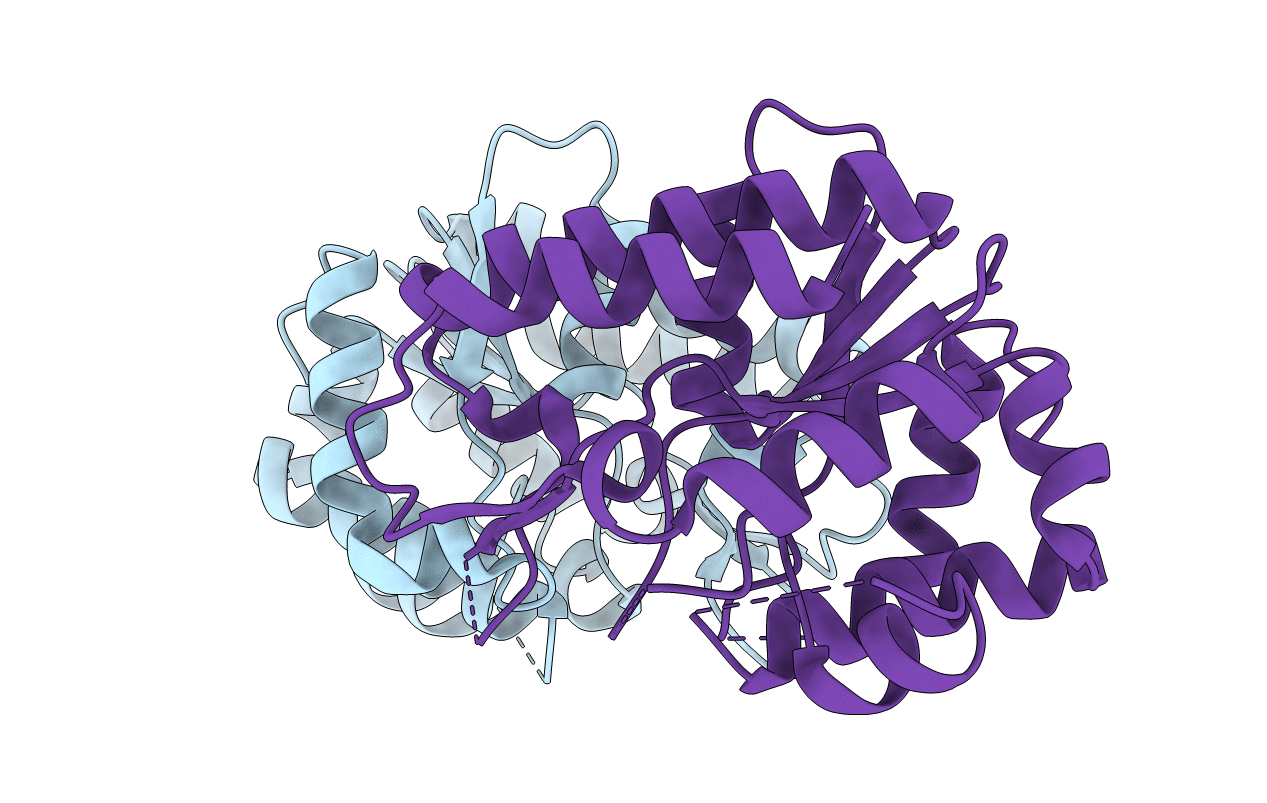
Resolution:
2.04 Å
R-Value Free:
0.23
R-Value Work:
0.22
R-Value Observed:
0.22
Space Group:
P 1 21 1
Macromolecular Entities
Structures with similar UniProt ID
Protein Blast
Polymer Type:polypeptide(L)
Molecule:Geobacter metallireducens SMUG1
Gene (Uniprot):Gmet_0095
Mutagens:G63P
Chain IDs:A, B
Chain Length:240
Number of Molecules:2
Biological Source:Geobacter metallireducens (strain GS-15 / ATCC 53774 / DSM 7210)
Primary Citation
Structural Basis of Substrate Specificity in Geobacter metallireducens SMUG1
Acs Chem.Biol. 11 1729 1736 (2016)
PMID: 27071000 DOI: 10.1021/acschembio.6b00164

Abstact

Base deamination is a common type of DNA damage that occurs in all organisms. DNA repair mechanisms are critical to maintain genome integrity, in which the base excision repair pathway plays an essential role. In the BER pathway, the uracil DNA glycosylase superfamily is responsible for removing the deaminated bases from DNA and generates apurinic/apyrimidinic (AP) sites. Geobacter metallireducens SMUG1 (GmeSMUG1) is an interesting family 3 enzyme in the UDG superfamily, with dual substrate specificities for DNA with uracil or xanthine. In contrast, the mutant G63P of GmeSMUG1 has exclusive activity for uracil, while N58D is inactive for both substrates, as we have reported previously. However, the structural bases for these substrate specificities are not well understood. In this study, we solved a series of crystal structures of WT and mutants of GmeSMUG1 at relatively high resolutions. These structures provide insight on the molecular mechanism of xanthine recognition for GmeSMUG1 and indicate that H210 plays a key role in xanthine recognition, which is in good agreement with the results of our EMSA and activity assays. More importantly, our mutant structures allow us to build models to rationalize our previous experimental observations of altered substrate activities of these mutants.

Legend

Protein

Chemical

Disease

Primary Citation of related structures
Feedback Form
Name
Email
Institute
Feedback