Deposition Date
2015-12-25
Release Date
2016-05-18
Last Version Date
2024-11-06
Entry Detail
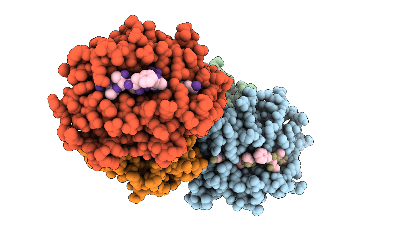
PDB ID:
5H94
Keywords:
Title:
Crystal structure of Swine MHC CLASSI for 1.48 angstroms
Biological Source:
Source Organism(s):
Sus scrofa (Taxon ID: 9823)
H1N1 swine influenza virus (Taxon ID: 36420)
H1N1 swine influenza virus (Taxon ID: 36420)
Expression System(s):
Method Details:
Experimental Method:
Resolution:
1.48 Å
R-Value Free:
0.19
R-Value Work:
0.14
R-Value Observed:
0.15
Space Group:
C 1 2 1