5H8T image
Deposition Date 2015-12-23
Release Date 2016-03-02
Last Version Date 2023-09-27
Entry Detail
PDB ID:
5H8T
Title:
Crystal structure of human cellular retinol binding protein 1 in complex with all-trans-retinol
Biological Source:
Source Organism:
Homo sapiens (Taxon ID: 9606)
Host Organism:
Method Details:
Experimental Method:
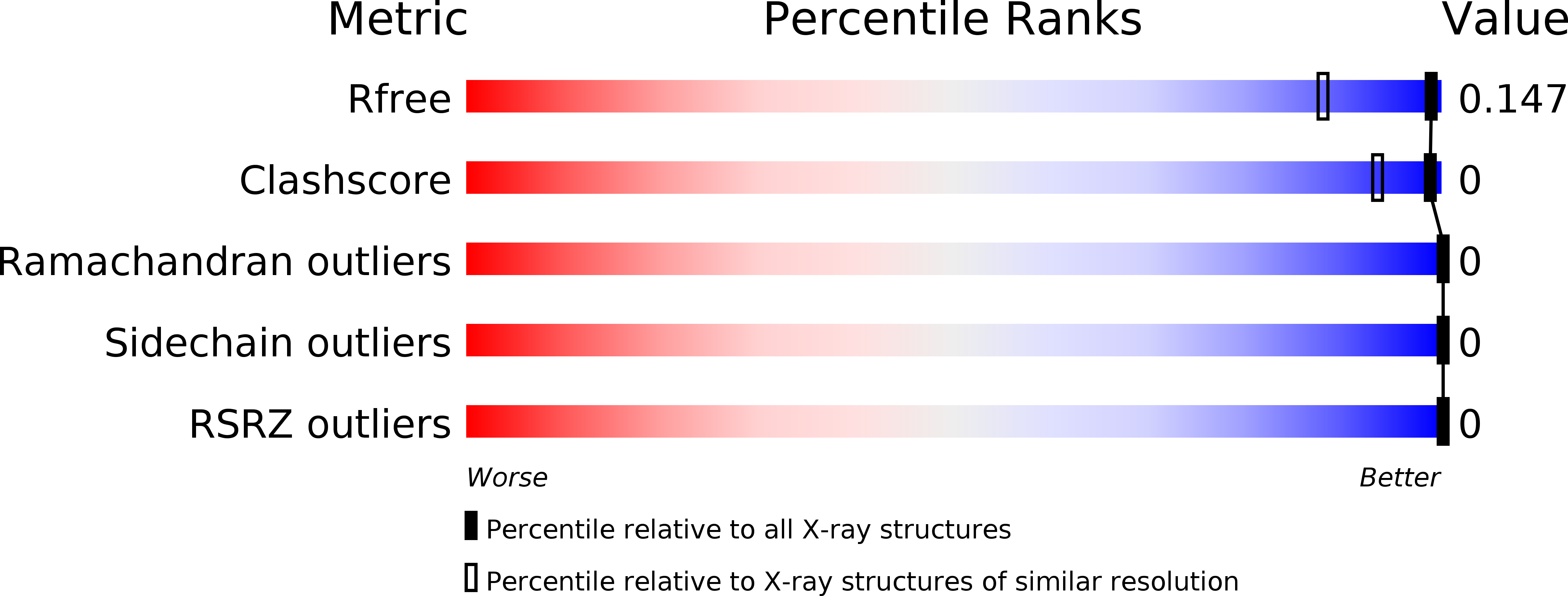
Resolution:
1.21 Å
R-Value Free:
0.14
R-Value Work:
0.11
R-Value Observed:
0.11
Space Group:
P 21 21 21
Macromolecular Entities
Structures with similar UniProt ID
Protein Blast
Polymer Type:polypeptide(L)
Molecule:Retinol-binding protein 1
Gene (Uniprot):RBP1
Chain IDs:A
Chain Length:140
Number of Molecules:1
Biological Source:Homo sapiens
Ligand Molecules
Primary Citation
Ligand Binding Induces Conformational Changes in Human Cellular Retinol-binding Protein 1 (CRBP1) Revealed by Atomic Resolution Crystal Structures.
J.Biol.Chem. 291 8528 8540 (2016)
PMID: 26900151 DOI: 10.1074/jbc.M116.714535

Abstact

Important in regulating the uptake, storage, and metabolism of retinoids, cellular retinol-binding protein 1 (CRBP1) is essential for trafficking vitamin A through the cytoplasm. However, the molecular details of ligand uptake and targeted release by CRBP1 remain unclear. Here we report the first structure of CRBP1 in a ligand-free form as well as ultra-high resolution structures of this protein bound to either all-trans-retinol or retinylamine, the latter a therapeutic retinoid that prevents light-induced retinal degeneration. Superpositioning of human apo- and holo-CRBP1 revealed major differences within segments surrounding the entrance to the retinoid-binding site. These included α-helix II and hairpin turns between β-strands βC-βD and βE-βF as well as several side chains, such as Phe-57, Tyr-60, and Ile-77, that change their orientations to accommodate the ligand. Additionally, we mapped hydrogen bond networks inside the retinoid-binding cavity and demonstrated their significance for the ligand affinity. Analyses of the crystallographic B-factors indicated several regions with higher backbone mobility in the apoprotein that became more rigid upon retinoid binding. This conformational flexibility of human apo-CRBP1 facilitates interaction with the ligands, whereas the more rigid holoprotein structure protects the labile retinoid moiety during vitamin A transport. These findings suggest a mechanism of induced fit upon ligand binding by mammalian cellular retinol-binding proteins.

Legend

Protein

Chemical

Disease

Primary Citation of related structures