5H6T image
Deposition Date 2016-11-15
Release Date 2017-03-15
Last Version Date 2023-11-08
Entry Detail
PDB ID:
5H6T
Keywords:
Title:
Crystal structure of Hydrazidase from Microbacterium sp. strain HM58-2
Biological Source:
Source Organism:
Host Organism:
Method Details:
Experimental Method:
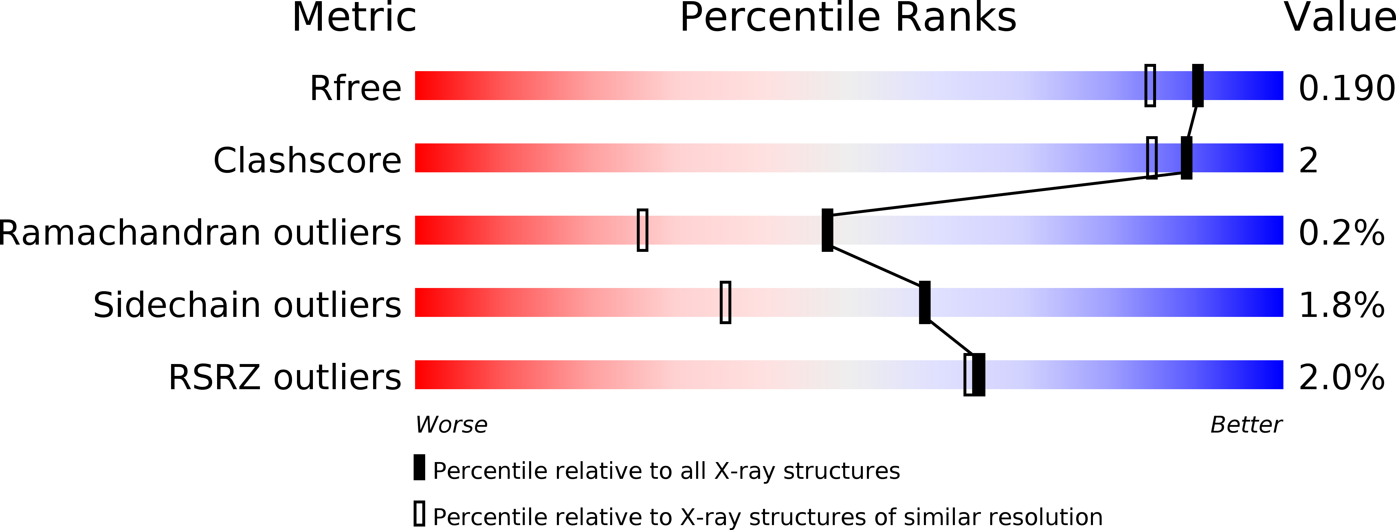
Resolution:
1.60 Å
R-Value Free:
0.17
R-Value Work:
0.16
R-Value Observed:
0.16
Space Group:
C 2 2 21
Macromolecular Entities
Polymer Type:polypeptide(L)
Molecule:Amidase
Chain IDs:A, B
Chain Length:481
Number of Molecules:2
Biological Source:Microbacterium sp. HM58-2
Primary Citation
Structural basis of the substrate recognition of hydrazidase isolated from Microbacterium sp. strain HM58-2, which catalyzes acylhydrazide compounds as its sole carbon source
Biochem. Biophys. Res. Commun. 482 1007 1012 (2017)
PMID: 27908731 DOI: 10.1016/j.bbrc.2016.11.148

Abstact

Hydrazidase was an enzyme that remained unidentified for a half century. However, recently, it was purified, and its encoding gene was cloned. Microbacterium sp. strain HM58-2 grows with acylhydrazides as its sole carbon source; it produces hydrazidase and degrades acylhydrazides to acetate and hydrazides. The bacterial hydrazidase belongs to the amidase signature enzyme family and contains a Ser-cisSer-Lys catalytic motif. The condensation of hydrazine and carbonic acid produces various hydrazides, some of which are raw materials for synthesizing pharmaceuticals and other useful chemicals. Although natural hydrazide compounds have been identified, the metabolic systems for hydrazides are not fully understood. Here, we report the crystal structure of hydrazidase from Microbacterium sp. strain HM58-2. The active site was revealed to consist of a Ser-cisSer-Lys catalytic triad, in which Ser179 forms a covalent bond with a carbonyl carbon of the substrate. 4-Hydroxybenzoic acid hydrazide bound to the S179A mutant, showing an oxyanion hole composed of the three backbone amide groups. Furthermore, H336 in the non-conserved region in the amidase family may define the substrate specificity, which was confirmed by mutation analysis. A wild-type apoenzyme structure revealed an unidentified molecule covalently bound to S179, representing a tetrahedral intermediate.

Legend

Protein

Chemical

Disease

Primary Citation of related structures