Deposition Date
2016-11-09
Release Date
2017-02-22
Last Version Date
2023-11-08
Entry Detail
PDB ID:
5H5W
Keywords:
Title:
Crystal structure of the flagellar cap protein FliD D2-D3 domains from Escherichia coli
Biological Source:
Source Organism(s):
Escherichia coli (Taxon ID: 562)
Expression System(s):
Method Details:
Experimental Method:
Resolution:
2.15 Å
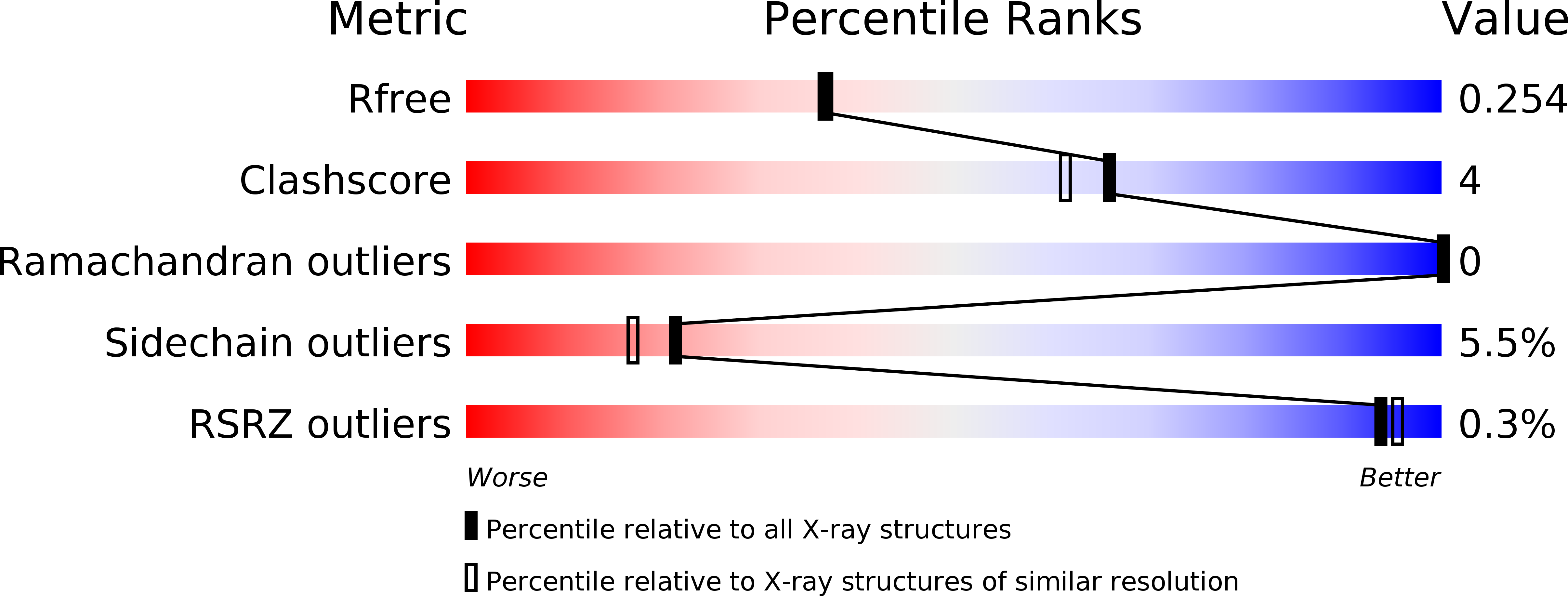
R-Value Free:
0.25
R-Value Work:
0.21
R-Value Observed:
0.21
Space Group:
P 6