Deposition Date
2016-11-04
Release Date
2017-05-03
Last Version Date
2024-11-20
Entry Detail
PDB ID:
5H58
Keywords:
Title:
Structural and dynamics studies of the TetR family protein, CprB from Streptomyces coelicolor in complex with its biological operator sequence
Biological Source:
Source Organism(s):
Streptomyces coelicolor A3(2) (Taxon ID: 100226)
Expression System(s):
Method Details:
Experimental Method:
Resolution:
3.99 Å
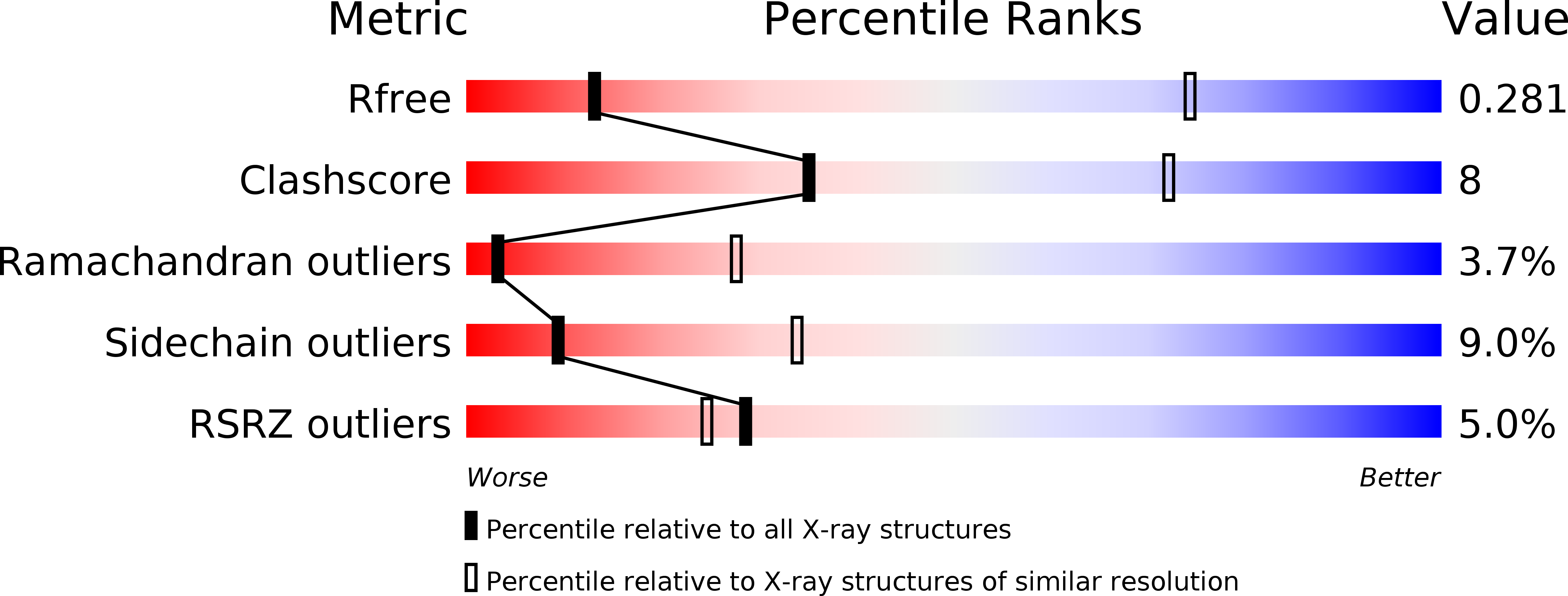
R-Value Free:
0.28
R-Value Work:
0.21
R-Value Observed:
0.22
Space Group:
P 32