
Deposition Date
2016-11-02
Release Date
2017-05-17
Last Version Date
2024-10-30
Entry Detail
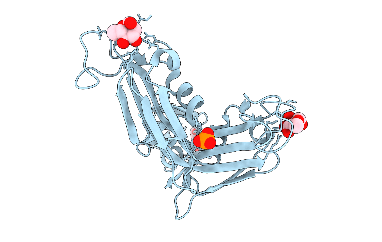
PDB ID:
5H4S
Keywords:
Title:
Crystal structure of a rhamnose-binding lectin SUL-I from the toxopneustid sea urchin Toxopneustes pileolus
Biological Source:
Source Organism(s):
Toxopneustes pileolus (Taxon ID: 39971)
Expression System(s):
Method Details:
Experimental Method:
Resolution:
1.80 Å
R-Value Free:
0.20
R-Value Work:
0.15
R-Value Observed:
0.15
Space Group:
C 1 2 1
