5H47 image
Deposition Date 2016-10-31
Release Date 2017-01-25
Last Version Date 2024-03-20
Entry Detail
PDB ID:
5H47
Title:
Crystal structure of AOL complexed with 2-MeSe-Fuc
Biological Source:
Source Organism:
Host Organism:
Method Details:
Experimental Method:
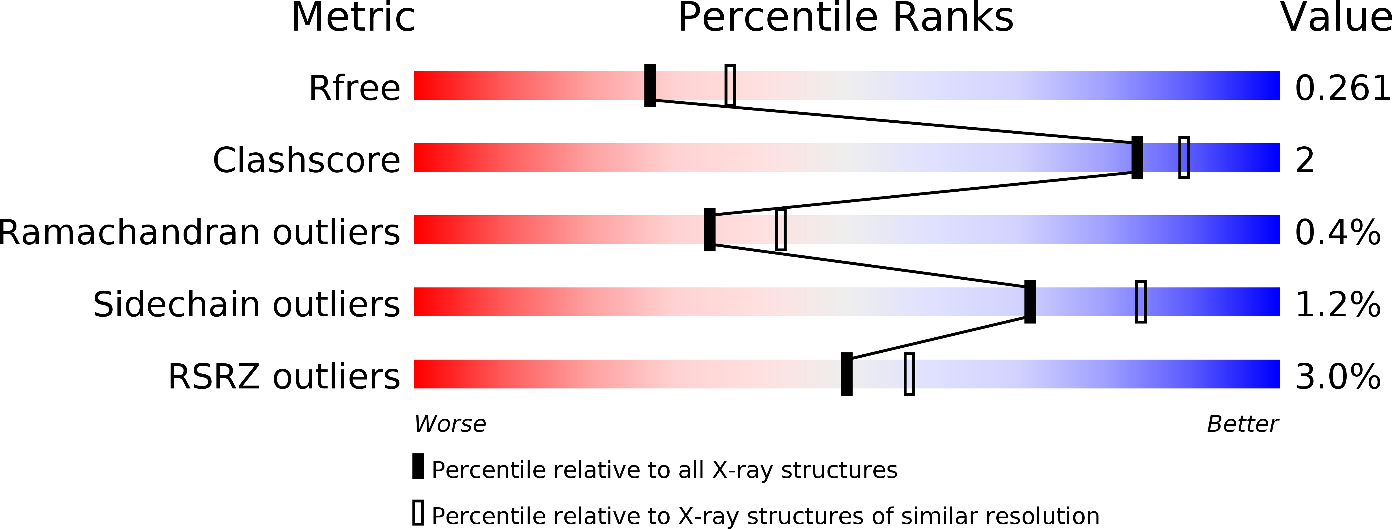
Resolution:
2.30 Å
R-Value Free:
0.25
R-Value Work:
0.21
R-Value Observed:
0.21
Space Group:
P 1 21 1
Macromolecular Entities
Structures with similar UniProt ID
Protein Blast
Polymer Type:polypeptide(L)
Molecule:Uncharacterized protein
Gene (Uniprot):fleA
Chain IDs:A, B, C, D, E, F, G, H, I, J, K, L
Chain Length:311
Number of Molecules:12
Biological Source:Aspergillus oryzae RIB40
Ligand Molecules
Primary Citation
Synthesis of seleno-fucose compounds and their application to the X-ray structural determination of carbohydrate-lectin complexes using single/multi-wavelength anomalous dispersion phasing
Bioorg. Med. Chem. 25 1132 1142 (2017)
PMID: 28041800 DOI: 10.1016/j.bmc.2016.12.021

Abstact

Selenium-incorporated fucoses (seleno-fucoses) differing in the position of the seleno-substituent were synthesized and applied to the X-ray structural determination of a carbohydrate-lectin complex using single/multi-wavelength anomalous dispersion (SAD/MAD) phasing. The hydroxyl groups at the C-1, -2, -3 and -4 position of fucose were individually substituted with a methylseleno group via a transacetalization reaction using MeSeCH2OBn or by an SN2 reaction with TolSe- equivalents to afford the corresponding MeSe-fucose. The three-dimensional structures of a fucose-binding lectin complexed with several of these MeSe-fucoses have been determined by SAD/MAD phasing by utilizing the diffraction of selenium in the bound MeSe-fucoses.

Legend

Protein

Chemical

Disease

Primary Citation of related structures