5H42 image
Deposition Date 2016-10-28
Release Date 2017-03-01
Last Version Date 2023-11-08
Entry Detail
PDB ID:
5H42
Keywords:
Title:
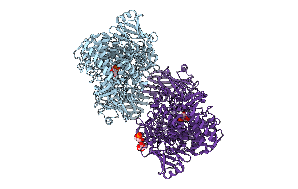
Crystal Structure of 1,2-beta-oligoglucan phosphorylase from Lachnoclostridium phytofermentans in complex with alpha-d-glucose-1-phosphate
Biological Source:
Source Organism:
Method Details:
Experimental Method:
Resolution:
2.10 Å
R-Value Free:
0.23
R-Value Work:
0.19
R-Value Observed:
0.19
Space Group:
P 1 21 1
Macromolecular Entities
Polymer Type:polypeptide(L)
Molecule:Uncharacterized protein
Gene (Uniprot):Cphy_0694
Chain IDs:A, B
Chain Length:1122
Number of Molecules:2
Biological Source:Clostridium phytofermentans ISDg
Primary Citation
Mechanistic insight into the substrate specificity of 1,2-beta-oligoglucan phosphorylase from Lachnoclostridium phytofermentans
Sci Rep 7 42671 42671 (2017)
PMID: 28198470 DOI: 10.1038/srep42671

Abstact

Glycoside phosphorylases catalyze the phosphorolysis of oligosaccharides into sugar phosphates. Recently, we found a novel phosphorylase acting on β-1,2-glucooligosaccharides with degrees of polymerization of 3 or more (1,2-β-oligoglucan phosphorylase, SOGP) in glycoside hydrolase family (GH) 94. Here, we characterized SOGP from Lachnoclostridium phytofermentans (LpSOGP) and determined its crystal structure. LpSOGP is a monomeric enzyme that contains a unique β-sandwich domain (Ndom1) at its N-terminus. Unlike the dimeric GH94 enzymes possessing catalytic pockets at their dimer interface, LpSOGP has a catalytic pocket between Ndom1 and the catalytic domain. In the complex structure of LpSOGP with sophorose, sophorose binds at subsites +1 to +2. Notably, the Glc moiety at subsite +1 is flipped compared with the corresponding ligands in other GH94 enzymes. This inversion suggests the great distortion of the glycosidic bond between subsites -1 and +1, which is likely unfavorable for substrate binding. Compensation for this disadvantage at subsite +2 can be accounted for by the small distortion of the glycosidic bond in the sophorose molecule. Therefore, the binding mode at subsites +1 and +2 defines the substrate specificity of LpSOGP, which provides mechanistic insights into the substrate specificity of a phosphorylase acting on β-1,2-glucooligosaccharides.

Legend

Protein

Chemical

Disease

Primary Citation of related structures