Deposition Date
2016-10-26
Release Date
2017-08-02
Last Version Date
2023-11-08
Entry Detail
PDB ID:
5H3R
Keywords:
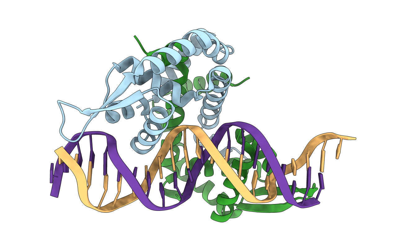
Title:
Crystal Structure of mutant MarR C80S from E.coli complexed with operator DNA
Biological Source:
Source Organism(s):
Escherichia coli (Taxon ID: 83333)
synthetic construct (Taxon ID: 32630)
synthetic construct (Taxon ID: 32630)
Expression System(s):
Method Details:
Experimental Method:
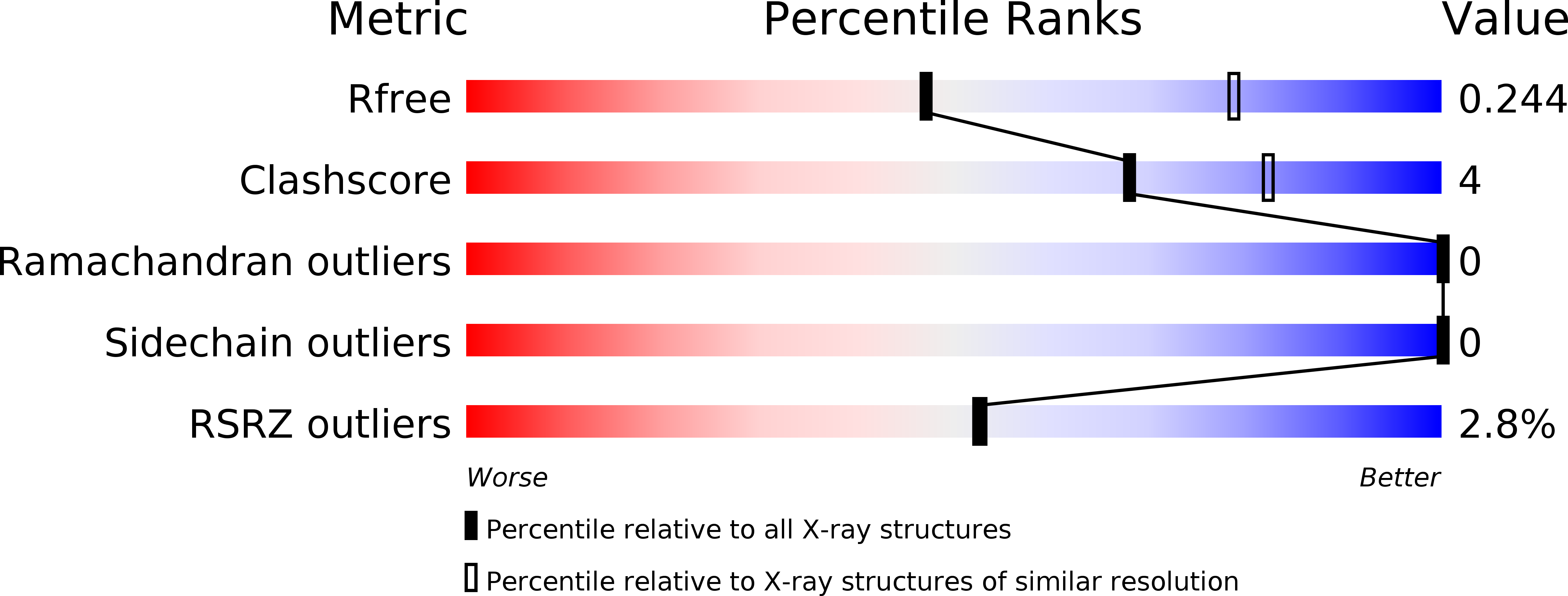
Resolution:
2.67 Å
R-Value Free:
0.24
R-Value Work:
0.19
R-Value Observed:
0.20
Space Group:
P 61