Deposition Date
2016-10-15
Release Date
2017-03-22
Last Version Date
2024-10-23
Entry Detail
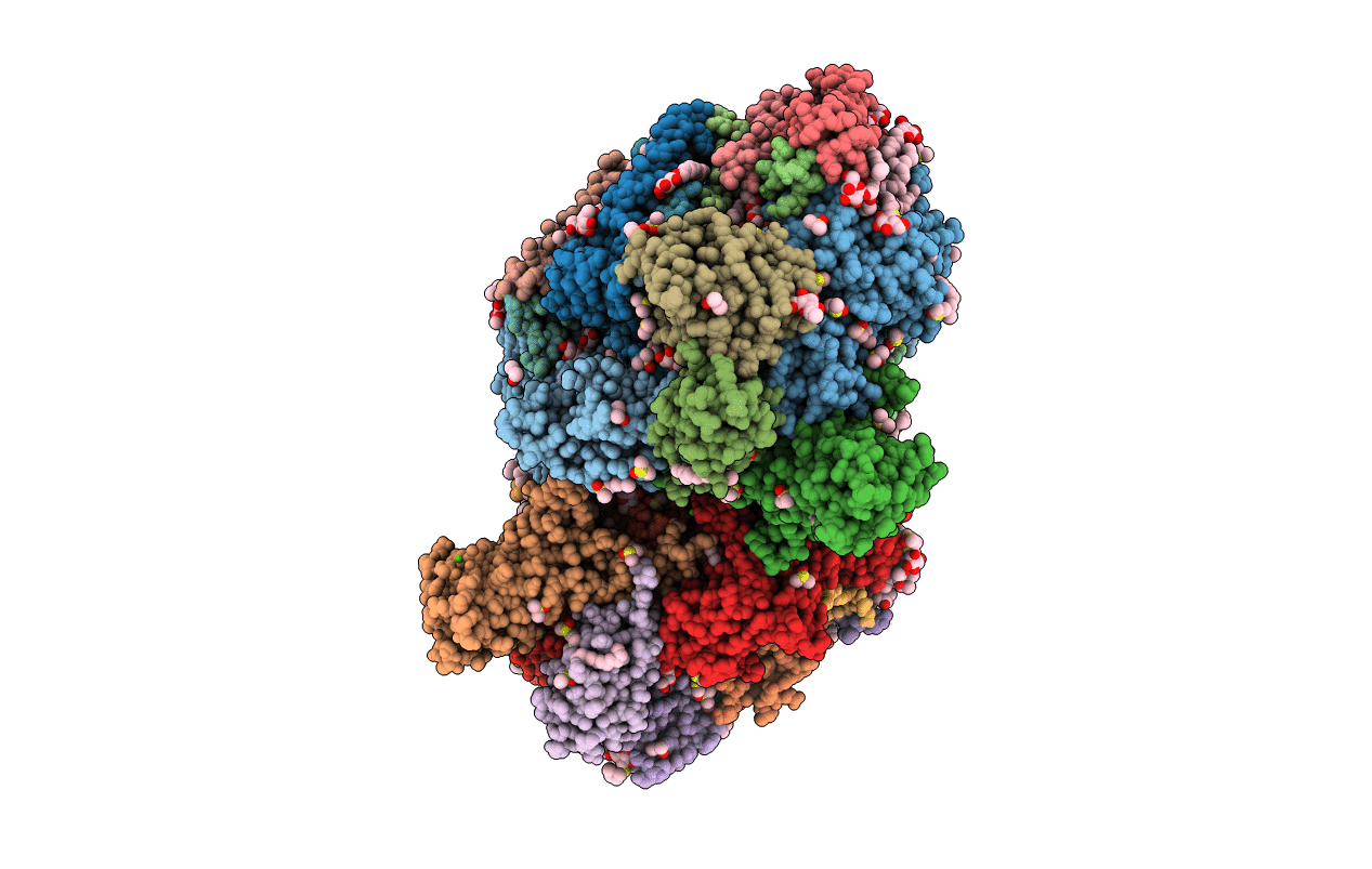
PDB ID:
5H2F
Keywords:
Title:
Crystal structure of the PsbM-deletion mutant of photosystem II
Biological Source:
Source Organism(s):
Thermosynechococcus elongatus (strain BP-1) (Taxon ID: 197221)
Method Details:
Experimental Method:
Resolution:
2.20 Å
R-Value Free:
0.22
R-Value Work:
0.17
R-Value Observed:
0.17
Space Group:
P 21 21 21