5H23 image
Deposition Date 2016-10-14
Release Date 2018-03-14
Last Version Date 2023-11-08
Entry Detail
PDB ID:
5H23
Keywords:
Title:
Crystal structure of Chikungunya virus capsid protein
Biological Source:
Source Organism:
Host Organism:
Method Details:
Experimental Method:
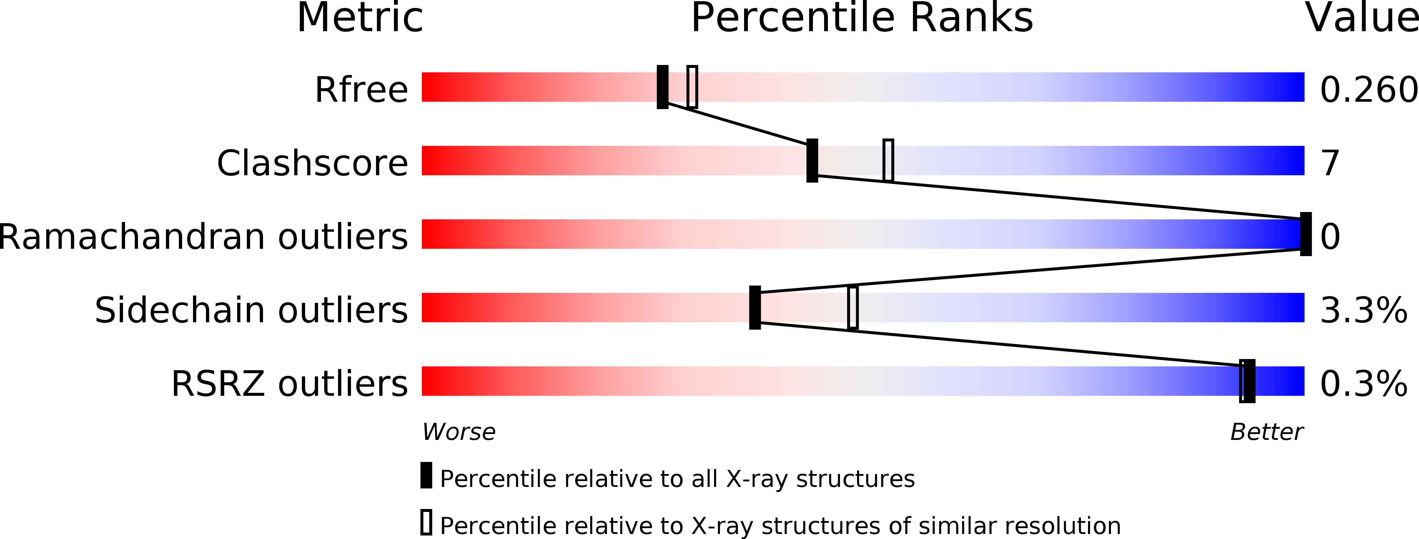
Resolution:
2.20 Å
R-Value Free:
0.26
R-Value Work:
0.17
R-Value Observed:
0.18
Space Group:
P 1
Macromolecular Entities
Polymer Type:polypeptide(L)
Molecule:Capsid Protein
Chain IDs:A, B
Chain Length:156
Number of Molecules:2
Biological Source:Chikungunya virus
Primary Citation
Structure-function insights into chikungunya virus capsid protein: Small molecules targeting capsid hydrophobic pocket.
Virology 515 223 234 (2018)
PMID: 29306785 DOI: 10.1016/j.virol.2017.12.020

Abstact

The crystal structure of chikungunya (CHIKV) virus capsid protease domain has been determined at 2.2Å. Structure reveals a chymotrypsin-like protease fold with a conserved hydrophobic pocket in CHIKV capsid protein (CP) for interaction with the cytoplasmic tail of E2 (cdE2) similar to the capsid protein of other alphaviruses. Molecular contacts between CP-cdE2 were determined by fitting structures of CHIKV CP and cdE2 into the cryo-EM map of Venezuelan equine encephalitis virus (VEEV). Binding of (S)-(+)-Mandelic acid (MDA) and Ethyl 3-aminobenzoate (EAB) to the hydrophobic pocket of CP was evaluated by molecular docking. Surface plasmon resonance (SPR) and fluorescence spectroscopy experiments confirmed MDA and EAB binding to the CP. The binding constants (KD) obtained from SPR for MDA and EAB were 1.2 × 10⁻³ M and 0.2 × 10⁻⁹ M, respectively. This study adds to the understanding of chikungunya virus structural proteins and may serve as the basis for antiviral development against chikungunya disease.

Legend

Protein

Chemical

Disease

Primary Citation of related structures