5H18 image
Deposition Date 2016-10-08
Release Date 2017-09-27
Last Version Date 2024-10-23
Entry Detail
PDB ID:
5H18
Keywords:
Title:
Crystal structure of catalytic domain of UGGT (UDP-glucose-bound form) from Thermomyces dupontii
Biological Source:
Source Organism:
Method Details:
Experimental Method:
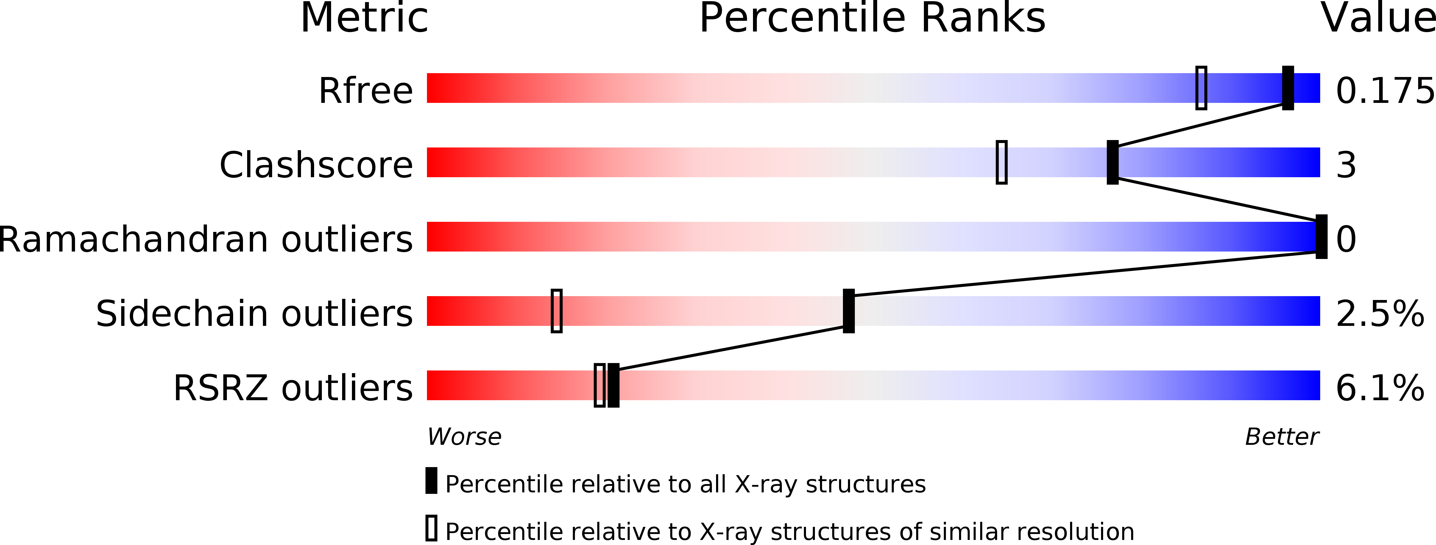
Resolution:
1.40 Å
R-Value Free:
0.17
R-Value Work:
0.14
R-Value Observed:
0.14
Space Group:
P 21 21 21
Macromolecular Entities
Structures with similar UniProt ID
Protein Blast
Polymer Type:polypeptide(L)
Molecule:UGGT
Chain IDs:A
Chain Length:293
Number of Molecules:1
Biological Source:Thermomyces dupontii
Primary Citation
Visualisation of a flexible modular structure of the ER folding-sensor enzyme UGGT.
Sci Rep 7 12142 12142 (2017)
PMID: 28939828 DOI: 10.1038/s41598-017-12283-w

Abstact

In the endoplasmic reticulum (ER), a protein quality control system facilitates the efficient folding of newly synthesised proteins. In this system, a series of N-linked glycan intermediates displayed on the protein surface serve as quality tags. The ER folding-sensor enzyme UDP-glucose:glycoprotein glucosyltransferase (UGGT) acts as a gatekeeper in the ER quality control system by specifically catalysing monoglucosylation onto incompletely folded glycoproteins, thereby enabling them to interact with lectin-chaperone complexes. Here we characterise the dynamic structure of this enzyme. Our crystallographic data demonstrate that the sensor region is composed of four thioredoxin-like domains followed by a β-rich domain, which are arranged into a C-shaped structure with a large central cavity, while the C-terminal catalytic domain undergoes a ligand-dependent conformational alteration. Furthermore, small-angle X-ray scattering, cryo-electron microscopy and high-speed atomic force microscopy have demonstrated that UGGT has a flexible modular structure in which the smaller catalytic domain is tethered to the larger folding-sensor region with variable spatial arrangements. These findings provide structural insights into the working mechanism whereby UGGT operates as a folding-sensor against a variety of glycoprotein substrates through its flexible modular structure possessing extended hydrophobic surfaces for the recognition of unfolded substrates.

Legend

Protein

Chemical

Disease

Primary Citation of related structures
Feedback Form
Name
Email
Institute
Feedback