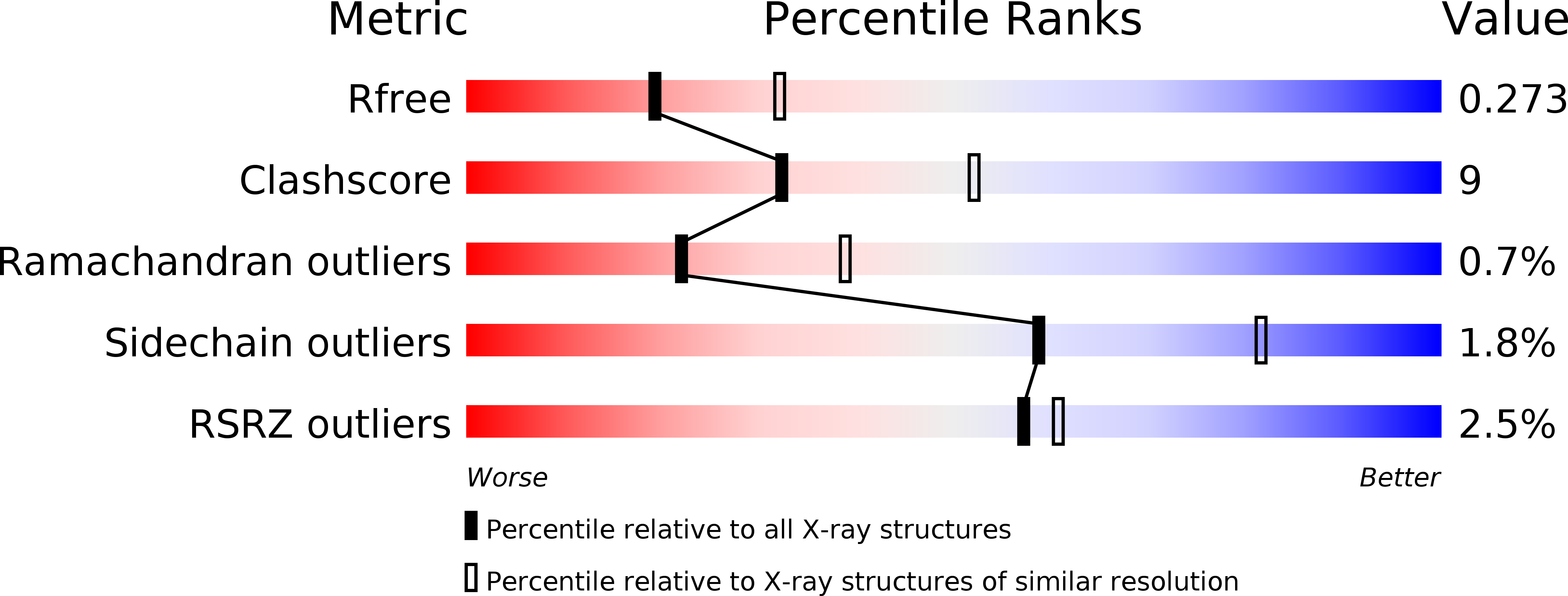
5H12 image
Deposition Date 2016-10-07
Release Date 2017-01-25
Last Version Date 2024-11-06
Entry Detail
PDB ID:
5H12
Keywords:
Title:
Crystal structure of Deep Vent DNA Polymerase
Biological Source:
Source Organism:
Pyrococcus (Taxon ID: 2260)
Host Organism:
Method Details:
Experimental Method:
Resolution:
2.50 Å
R-Value Free:
0.27
R-Value Work:
0.21
R-Value Observed:
0.21
Space Group:
P 21 21 21
Macromolecular Entities
Structures with similar UniProt ID
Protein Blast
Polymer Type:polypeptide(L)
Molecule:Deep Vent DNA Polymerase
Chain IDs:A
Chain Length:775
Number of Molecules:1
Biological Source:Pyrococcus
Primary Citation
Crystal structure of Deep Vent DNA polymerase.
Biochem. Biophys. Res. Commun. 483 52 57 (2017)
PMID: 28063932 DOI: 10.1016/j.bbrc.2017.01.007

Abstact

DNA polymerases are useful tools in various biochemical experiments. We have focused on the DNA polymerases involved in DNA replication including the unnatural base pair between 7-(2-thienyl)imidazo[4,5-b]pyridine (Ds) and 2-nitro-4-propynylpyrrole (Px). Many reports have described the different combinations between unnatural base pairs and DNA polymerases. As an example, for the replication of the Ds-Px pair, Deep Vent DNA polymerase exhibits high efficiency and fidelity, but Taq DNA polymerase shows much lower efficiency and fidelity. In the present study, we determined the crystal structure of Deep Vent DNA polymerase in the apo form at 2.5 Å resolution. Using this structure, we constructed structural models of Deep Vent DNA polymerase complexes with DNA containing an unnatural or natural base in the replication position. The models revealed that the unnatural Ds base in the template-strand DNA clashes with the side-chain oxygen of Thr664 in Taq DNA polymerase, but not in Deep Vent DNA polymerase.

Legend

Protein

Chemical

Disease

Primary Citation of related structures