Deposition Date
2016-09-03
Release Date
2017-04-05
Last Version Date
2023-11-08
Entry Detail
PDB ID:
5GV8
Keywords:
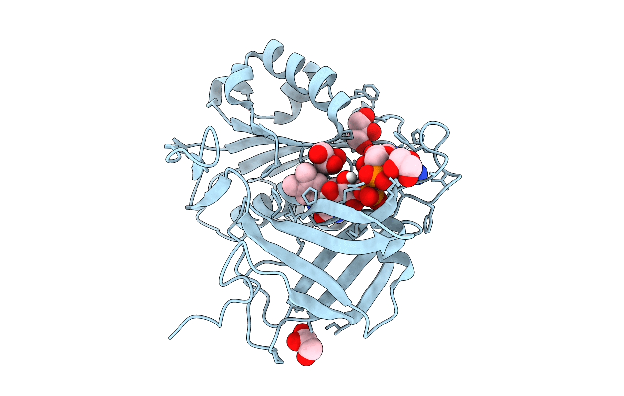
Title:
Structure of NADH-cytochrome b5 reductase refined with the multipolar atomic model at 0.78A
Biological Source:
Source Organism(s):
Sus scrofa (Taxon ID: 9823)
Expression System(s):
Method Details:
Experimental Method:
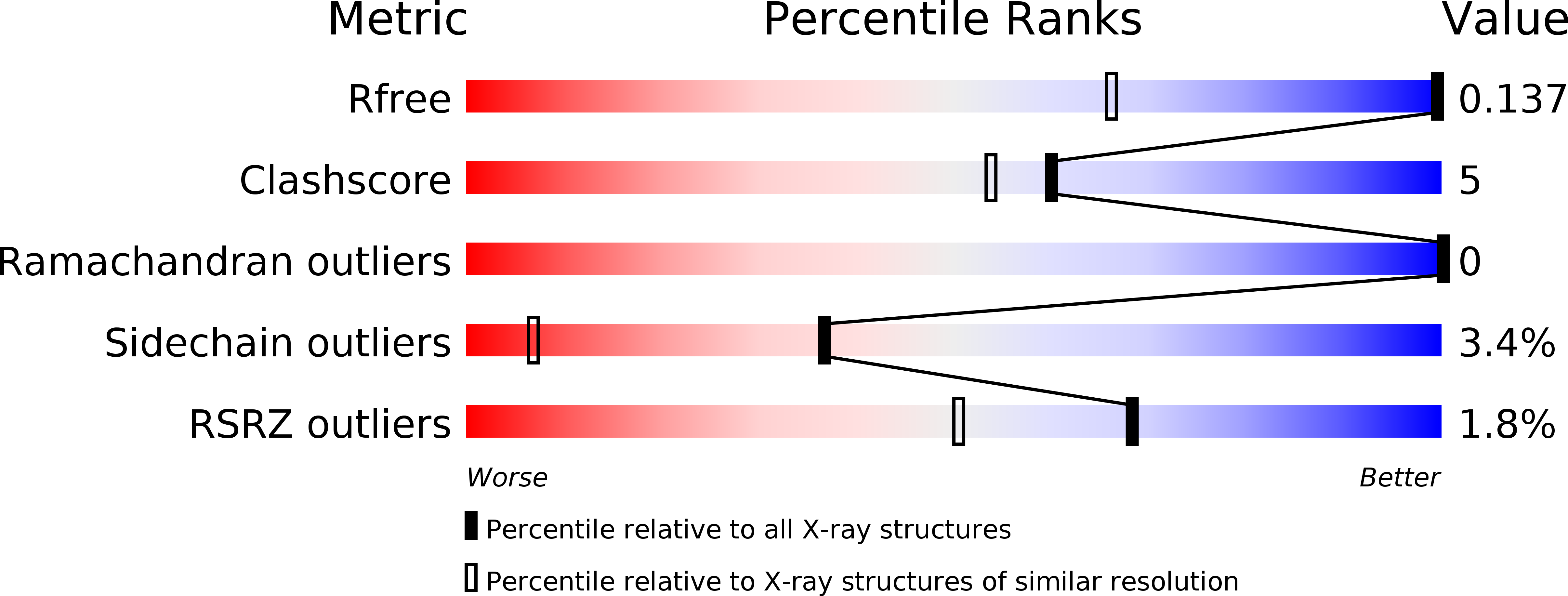
Resolution:
0.78 Å
R-Value Free:
0.14
R-Value Work:
0.12
Space Group:
P 21 21 21