5GU5 image
Deposition Date 2016-08-25
Release Date 2017-01-25
Last Version Date 2024-11-13
Entry Detail
PDB ID:
5GU5
Title:
Crystal structure of p24gamma2 GOLD domain determined by sulfur-SAD
Biological Source:
Source Organism:
Mus musculus (Taxon ID: 10090)
Host Organism:
Method Details:
Experimental Method:
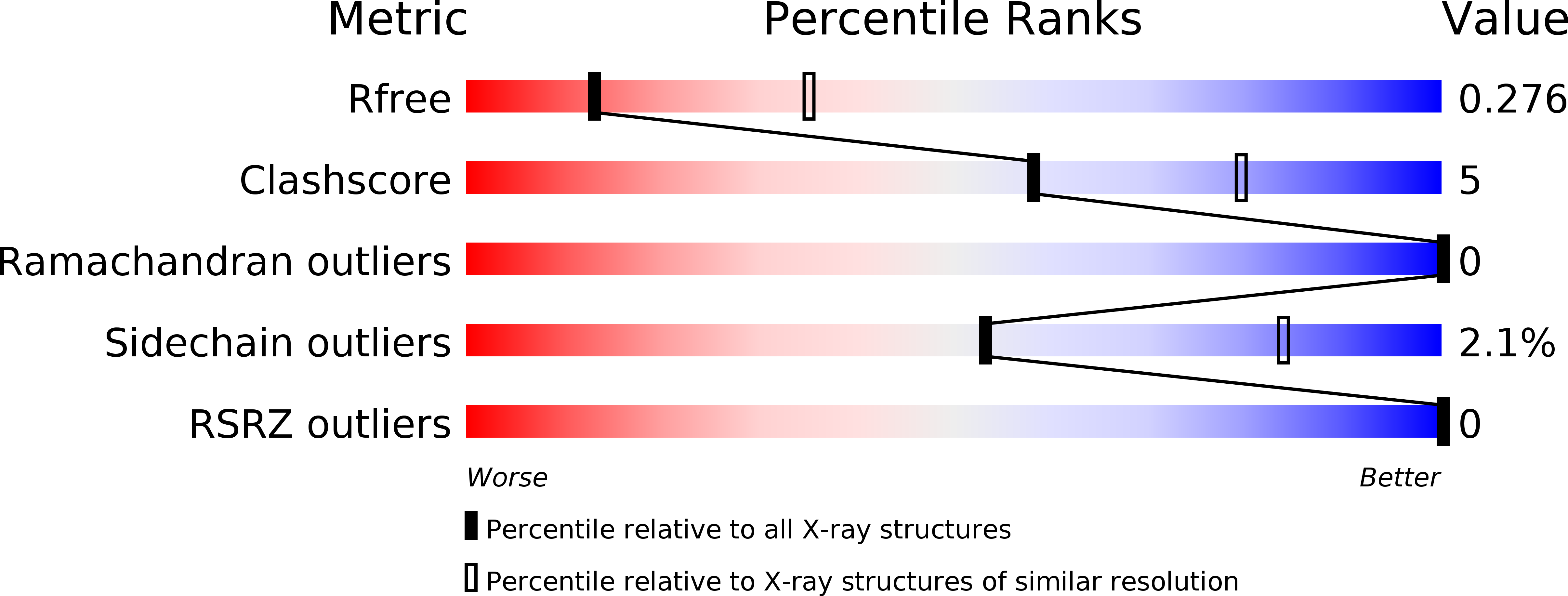
Resolution:
2.80 Å
R-Value Free:
0.27
R-Value Work:
0.22
R-Value Observed:
0.22
Space Group:
P 64 2 2
Macromolecular Entities
Structures with similar UniProt ID
Protein Blast
Polymer Type:polypeptide(L)
Molecule:Transmembrane emp24 domain-containing protein 5
Gene (Uniprot):Tmed5
Chain IDs:A
Chain Length:114
Number of Molecules:1
Biological Source:Mus musculus
Ligand Molecules
Primary Citation
Crystallographic analysis of murine p24 gamma 2 Golgi dynamics domain
Proteins 85 764 770 (2017)
PMID: 28066915 DOI: 10.1002/prot.25242

Abstact

The p24 family proteins form homo- and hetero-oligomeric complexes for efficient transport of cargo proteins from the endoplasmic reticulum to the Golgi apparatus. It consists of four subfamilies (p24α, p24β, p24γ, and p24δ). p24γ2 plays crucial roles in the selective transport of glycosylphosphatidylinositol-anchored proteins. Here, we determined the crystal structure of mouse p24γ2 Golgi dynamics (GOLD) domain at 2.8 Å resolution by the single anomalous diffraction method using intrinsic sulfur atoms. In spite of low sequence identity among p24 family proteins, p24γ2 GOLD domain assumes a β-sandwich fold, similar to that of p24β1 or p24δ1. An additional short α-helix is observed at the C-terminus of the p24γ2 GOLD domain. Intriguingly, p24γ2 GOLD domains crystallize as dimers, and dimer formation seems assisted by the short α-helix. Dimerization modes of GOLD domains are compared among p24 family proteins. Proteins 2017; 85:764-770. © 2016 Wiley Periodicals, Inc.

Legend

Protein

Chemical

Disease

Primary Citation of related structures
Feedback Form
Name
Email
Institute
Feedback