5GTP image
Entry Detail
PDB ID:
5GTP
Keywords:
Title:
The agonist-free structure of human PPARgamma ligand binding domain in the presence of the SRC-1 coactivator peptide
Biological Source:
Source Organism:
Host Organism:
PDB Version:
Deposition Date:
2016-08-23
Release Date:
2017-07-05
Method Details:
Experimental Method:
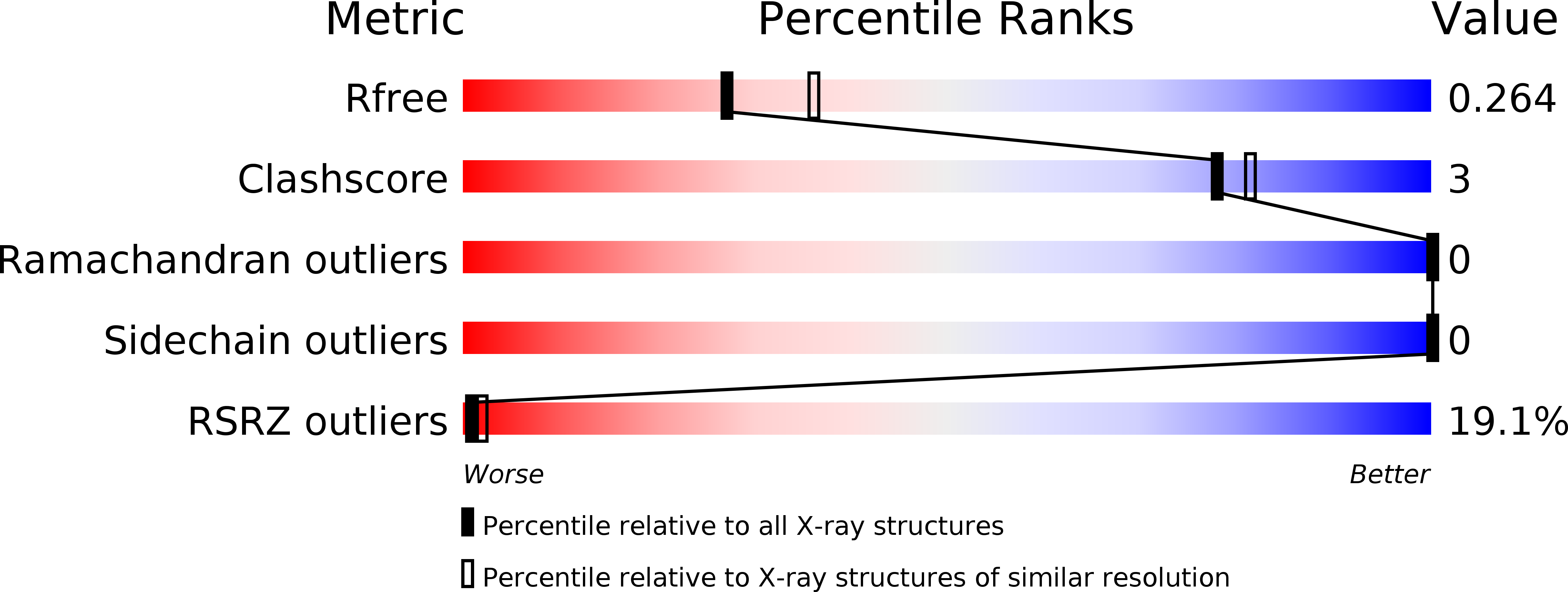
Resolution:
2.35 Å
R-Value Free:
0.25
R-Value Work:
0.21
R-Value Observed:
0.21
Space Group:
P 21 21 2
Macromolecular Entities
Polymer Type:polypeptide(L)
Description:Peroxisome proliferator-activated receptor gamma
Chain IDs:A
Chain Length:283
Number of Molecules:1
Biological Source:Homo sapiens
Polymer Type:polypeptide(L)
Description:Nuclear receptor coactivator 1
Chain IDs:B
Chain Length:16
Number of Molecules:1
Biological Source:Homo sapiens
Primary Citation
Structural basis for differential activities of enantiomeric PPAR gamma agonists: Binding of S35 to the alternate site.
Biochim. Biophys. Acta 1865 674 681 (2017)
PMID: 28342850 DOI: 10.1016/j.bbapap.2017.03.008

Abstact

Peroxisome proliferator-activated receptor γ (PPARγ) is a member of the nuclear receptor superfamily. It functions as a ligand-activated transcription factor and plays important roles in the regulation of adipocyte differentiation, type 2 diabetes mellitus, and inflammation. Many PPARγ agonists bind to the canonical ligand-binding pocket near the activation function-2 (AF-2) helix (i.e., helix H12) of the ligand-binding domain (LBD). More recently, an alternate ligand-binding site was identified in PPARγ LBD; it is located beside the Ω loop between the helices H2' and H3. We reported previously that the chirality of two optimized enantiomeric PPARγ ligands (S35 and R35) differentiates their PPARγ transcriptional activity, binding affinity, and inhibitory activity toward Cdk5 (cyclin-dependent kinase 5)-mediated phosphorylation of PPARγ at Ser245 (in PPARγ1 numbering; Ser273 in PPARγ2 numbering). S35 is a PPARγ phosphorylation inhibitor with promising glucose uptake potential, whereas R35 behaves as a potent conventional PPARγ agonist. To provide a structural basis for understanding the differential activities of these enantiomeric ligands, we have determined crystal structures of the PPARγ LBD in complex with either S35 or R35. S35 and R35 bind to the PPARγ LBD in significantly different manners. The partial agonist S35 occupies the alternate site near the Ω loop, whereas the full agonist R35 binds entirely to the canonical LBP. Alternate site binding of S35 affects the PPARγ transactivation and the inhibitory effect on PPARγ Ser245 phosphorylation. This study provides a useful platform for the development of a new generation of PPARγ ligands as anti-diabetic drug candidates.

Legend

Protein

Chemical

Disease

Primary Citation of related structures