5GT9 image
Deposition Date 2016-08-19
Release Date 2017-05-17
Last Version Date 2023-11-08
Entry Detail
PDB ID:
5GT9
Keywords:
Title:
The X-ray structure of 7beta-hydroxysteroid dehydrogenase
Biological Source:
Source Organism:
Method Details:
Experimental Method:
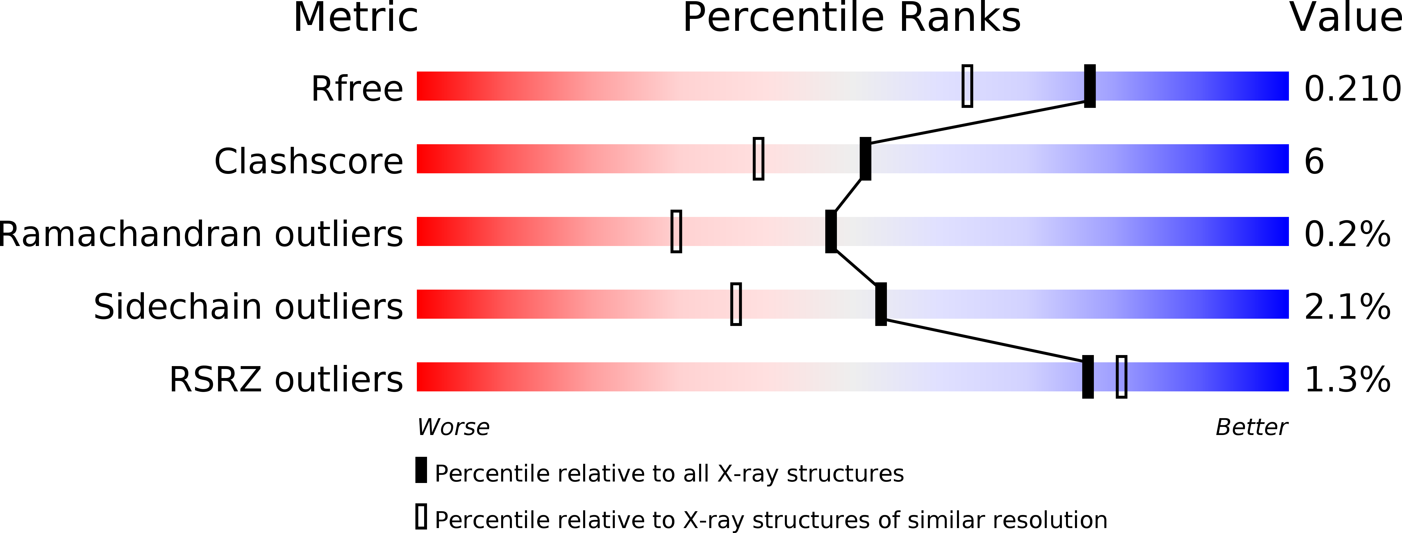
Resolution:
1.70 Å
R-Value Free:
0.20
R-Value Work:
0.16
R-Value Observed:
0.16
Space Group:
P 41 21 2
Macromolecular Entities
Polymer Type:polypeptide(L)
Molecule:Oxidoreductase, short chain dehydrogenase/reductase family protein
Gene (Uniprot):COLAER_02088
Chain IDs:A, B
Chain Length:263
Number of Molecules:2
Biological Source:Collinsella aerofaciens ATCC 25986
Ligand Molecules
Primary Citation
Structure of NADP(+)-bound 7 beta-hydroxysteroid dehydrogenase reveals two cofactor-binding modes
Acta Crystallogr F Struct Biol Commun 73 246 252 (2017)
PMID: 28471355 DOI: 10.1107/S2053230X17004460

Abstact

In mammals, bile acids/salts and their glycine and taurine conjugates are effectively recycled through enterohepatic circulation. 7β-Hydroxysteroid dehydrogenases (7β-HSDHs; EC 1.1.1.201), including that from the intestinal microbe Collinsella aerofaciens, catalyse the NADPH-dependent reversible oxidation of secondary bile-acid products to avoid potential toxicity. Here, the first structure of NADP+ bound to dimeric 7β-HSDH is presented. In one active site, NADP+ adopts a conventional binding mode similar to that displayed in related enzyme structures. However, in the other active site a unique binding mode is observed in which the orientation of the nicotinamide is different. Since 7β-HSDH has become an attractive target owing to the wide and important pharmaceutical use of its product ursodeoxycholic acid, this work provides a more detailed template to support rational protein engineering to improve the enzymatic activities of this useful biocatalyst, further improving the yield of ursodeoxycholic acid and its other applications.

Legend

Protein

Chemical

Disease

Primary Citation of related structures