5GPJ image
Deposition Date 2016-08-03
Release Date 2016-12-28
Last Version Date 2024-10-16
Entry Detail
PDB ID:
5GPJ
Keywords:
Title:
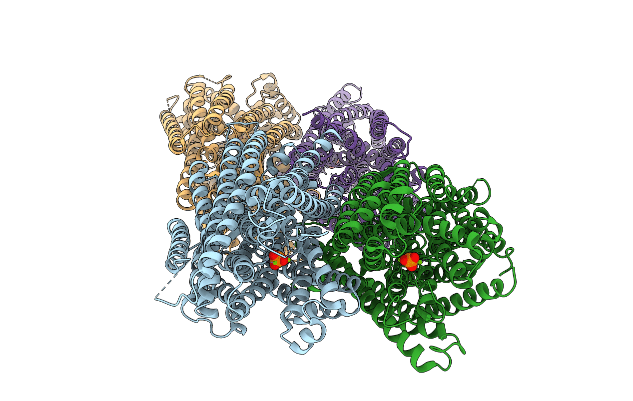
Crystal Structure of Proton-Pumping Pyrophosphatase
Biological Source:
Source Organism:
Method Details:
Experimental Method:
Resolution:
3.50 Å
R-Value Free:
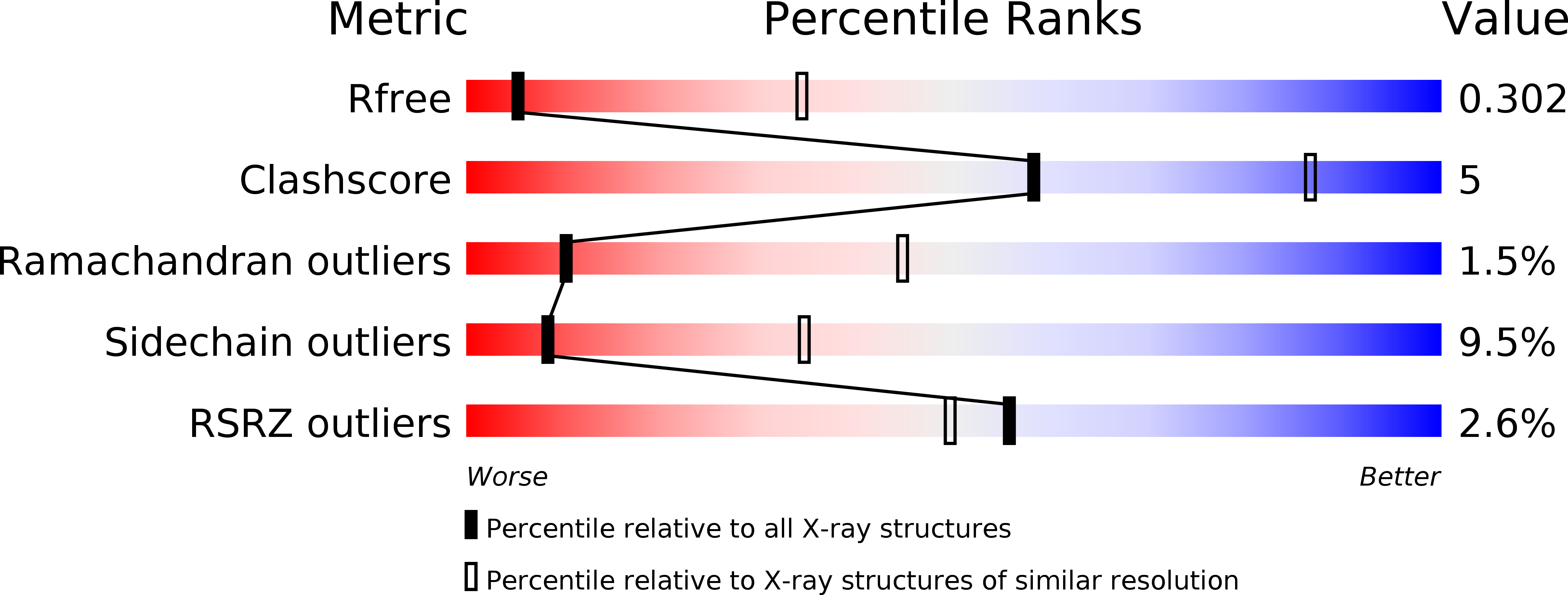
0.30
R-Value Work:
0.22
R-Value Observed:
0.22
Space Group:
C 1 2 1
Macromolecular Entities
Polymer Type:polypeptide(L)
Molecule:Pyrophosphate-energized vacuolar membrane proton pump
Chain IDs:A, B, C, D
Chain Length:774
Number of Molecules:4
Biological Source:Vigna radiata var. radiata
Primary Citation
Membrane pyrophosphatases from Thermotoga maritima and Vigna radiata suggest a conserved coupling mechanism
Nat Commun 7 13596 13596 (2016)
PMID: 27922000 DOI: 10.1038/ncomms13596

Abstact

Membrane-bound pyrophosphatases (M-PPases), which couple proton/sodium ion transport to pyrophosphate synthesis/hydrolysis, are important in abiotic stress resistance and in the infectivity of protozoan parasites. Here, three M-PPase structures in different catalytic states show that closure of the substrate-binding pocket by helices 5-6 affects helix 13 in the dimer interface and causes helix 12 to move down. This springs a 'molecular mousetrap', repositioning a conserved aspartate and activating the nucleophilic water. Corkscrew motion at helices 6 and 16 rearranges the key ionic gate residues and leads to ion pumping. The pumped ion is above the ion gate in one of the ion-bound structures, but below it in the other. Electrometric measurements show a single-turnover event with a non-hydrolysable inhibitor, supporting our model that ion pumping precedes hydrolysis. We propose a complete catalytic cycle for both proton and sodium-pumping M-PPases, and one that also explains the basis for ion specificity.

Legend

Protein

Chemical

Disease

Primary Citation of related structures