5GNL image
Deposition Date 2016-07-22
Release Date 2017-05-17
Last Version Date 2023-11-08
Entry Detail
PDB ID:
5GNL
Keywords:
Title:
Cytochrome P450 Vdh (CYP107BR1) F106V mutant
Biological Source:
Source Organism:
Host Organism:
Method Details:
Experimental Method:
Resolution:
1.95 Å
R-Value Free:
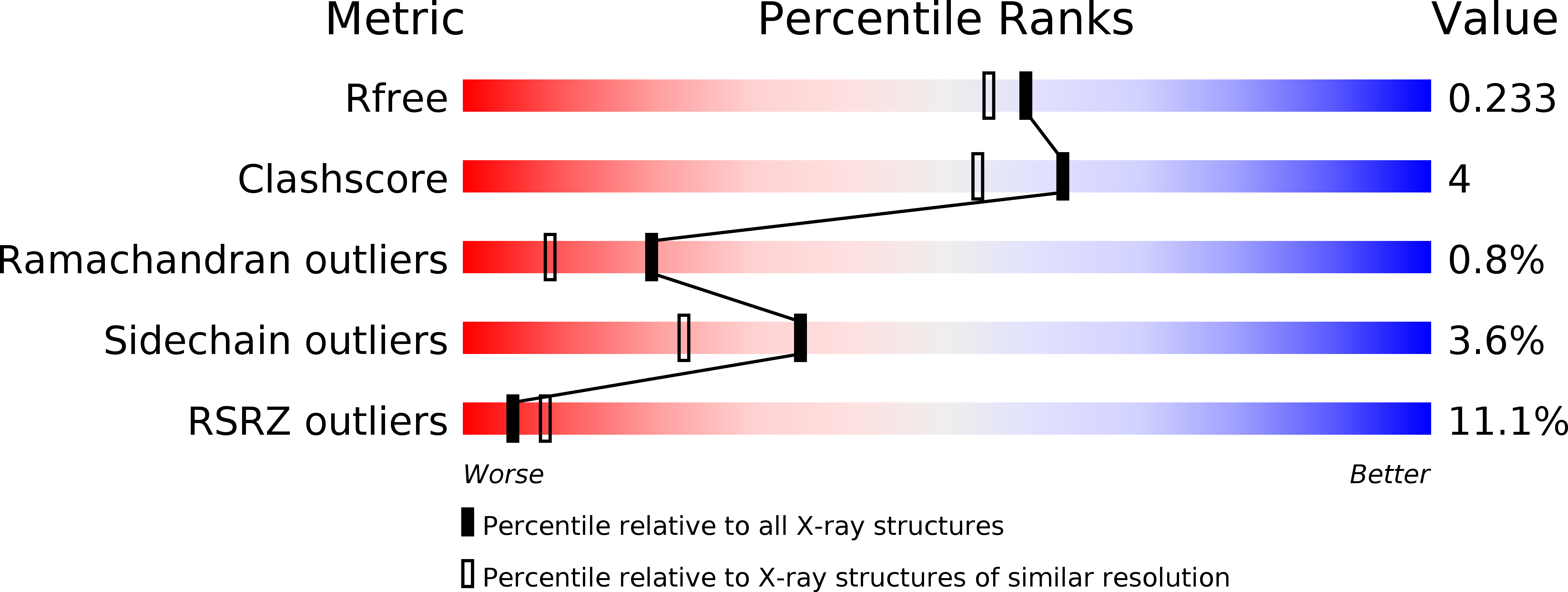
0.23
R-Value Work:
0.19
R-Value Observed:
0.20
Space Group:
P 31
Macromolecular Entities
Polymer Type:polypeptide(L)
Molecule:Vitamin D(3) 25-hydroxylase
Gene (Uniprot):vdh
Mutations:F106V
Chain IDs:A
Chain Length:411
Number of Molecules:1
Biological Source:Pseudonocardia autotrophica
Primary Citation
Structural insights into the mechanism of the drastic changes in enzymatic activity of the cytochrome P450 vitamin D3 hydroxylase (CYP107BR1) caused by a mutation distant from the active site
Acta Crystallogr F Struct Biol Commun 73 266 275 (2017)
PMID: 28471358 DOI: 10.1107/S2053230X17004782

Abstact

Cytochromes P450 (P450s) are haem-containing enzymes that catalyze medically and industrially important oxidative reactions, and many P450s have been subjected to directed evolution and site-directed mutagenesis to improve their activity and substrate specificity. Nonetheless, in most cases the mechanism that leads to drastic changes in specific activity after the introduction of an amino-acid substitution distant from the active-site pocket is unclear. Here, two crystal structures of inactive mutants of the P450 vitamin D3 hydroxylase (Vdh), Vdh-F106V and Vdh-L348M, which were obtained in the course of protein-engineering experiments on Vdh, are reported. The overall structures of these mutants show an open conformation similar to that of wild-type Vdh (Vdh-WT), whereas a rearrangement of the common main-chain hydrogen bonds is observed in the CD-loop (residues 102-106), resulting in a more compactly folded CD-loop relative to that of Vdh-WT. The previously reported structures of Vdh-WT and of the highly active Vdh-T107A and Vdh-K1 mutants have a more stretched CD-loop, with partial formation of 310-helix-type hydrogen bonds, both in the open and closed states. Molecular-dynamics simulations also showed that the frequency of the 310-helix is significantly reduced in Vdh-F106V and Vdh-L348M. The closed conformation is crucial for substrate and ferredoxin binding to initiate the catalytic reaction of Vdh. Therefore, it is implied that the small local structural changes observed in this study might disrupt the conformational transition from the open to the closed state, thereby leading to a complete loss of vitamin D3 hydroxylase activity.

Legend

Protein

Chemical

Disease

Primary Citation of related structures