5GLF image
Deposition Date 2016-07-11
Release Date 2016-11-09
Last Version Date 2023-11-08
Entry Detail
PDB ID:
5GLF
Title:
Structural insights into the interaction of p97 N-terminal domain and SHP motif in Derlin-1 rhomboid pseudoprotease
Biological Source:
Source Organism:
Homo sapiens (Taxon ID: 9606)
Host Organism:
Method Details:
Experimental Method:
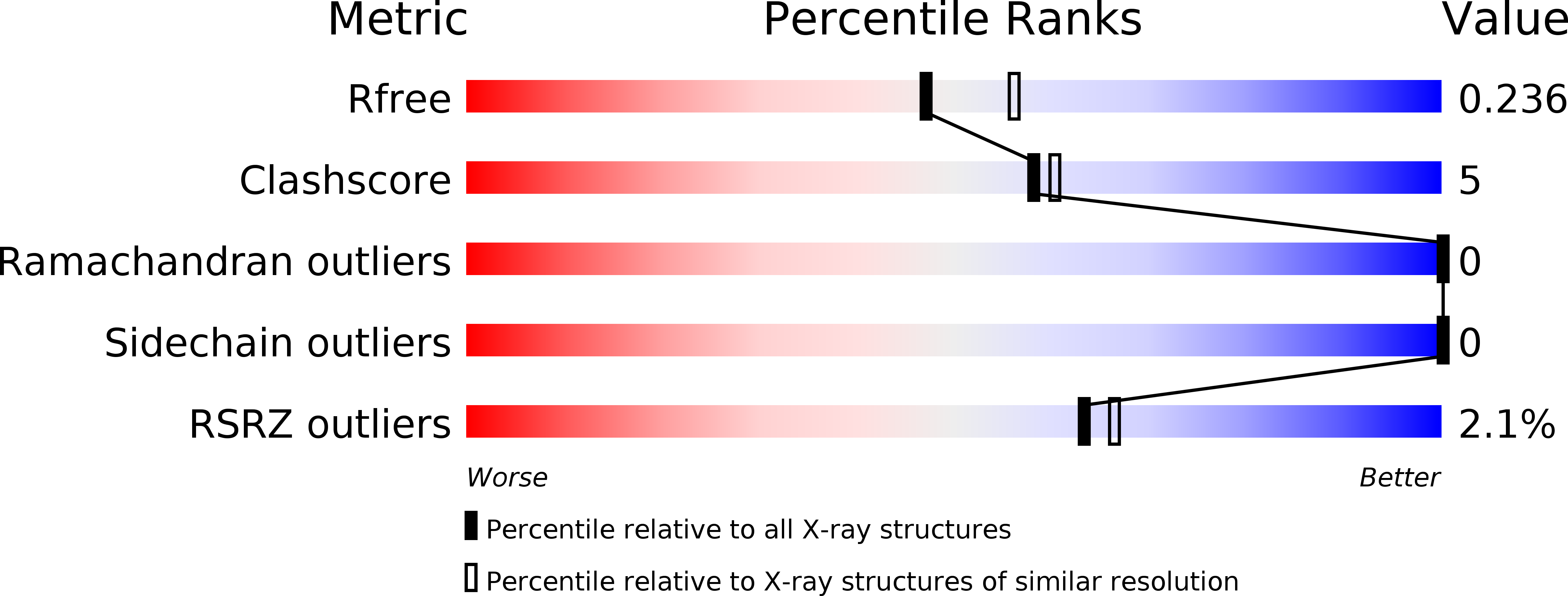
Resolution:
2.25 Å
R-Value Free:
0.23
R-Value Work:
0.18
R-Value Observed:
0.19
Space Group:
P 21 21 21
Macromolecular Entities
Structures with similar UniProt ID
Protein Blast
Polymer Type:polypeptide(L)
Molecule:Transitional endoplasmic reticulum ATPase
Gene (Uniprot):VCP
Chain IDs:A, C, E, G
Chain Length:184
Number of Molecules:4
Biological Source:Homo sapiens
Structures with similar UniProt ID
Protein Blast
Polymer Type:polypeptide(L)
Molecule:Derlin-1
Gene (Uniprot):DERL1
Chain IDs:B, D, F, H
Chain Length:12
Number of Molecules:4
Biological Source:Homo sapiens
Primary Citation
Structural insights into the interaction of human p97 N-terminal domain and SHP motif in Derlin-1 rhomboid pseudoprotease.
FEBS Lett. 590 4402 4413 (2016)
PMID: 27714797 DOI: 10.1002/1873-3468.12447

Abstact

The interaction of the rhomboid pseudoprotease Derlin-1 and p97 is crucial for the retrotranslocation of polyubiquitinated substrates in the endoplasmic reticulum-associated degradation pathway. We report a 2.25 Å resolution structure of the p97 N-terminal domain (p97N) in complex with the Derlin-1 SHP motif. Remarkably, the SHP motif adopts a short, antiparallel β-strand that interacts with the β-sheet of p97N-a site distinct from that to which most p97 adaptor proteins bind. Mutational and biochemical analyses contributed to defining the specific interaction, demonstrating the importance of a highly conserved binding pocket on p97N and a signature motif on SHP. Our findings may also provide insights into the interactions between other SHP-containing proteins and p97N.

Legend

Protein

Chemical

Disease

Primary Citation of related structures